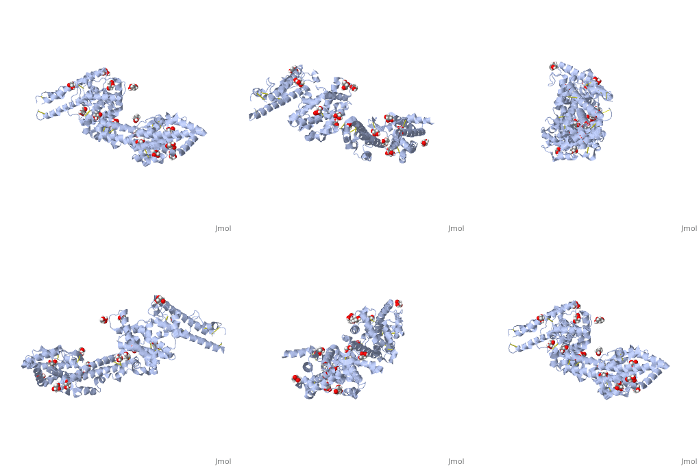
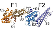
The EBL family members, including PfEBA-140, are made up of two regions, region II (RII) (shown to the right) and region VI.[2] Region II is responsible for receptor binding in all EBL family members.[2] RII is composed of , F1 (purple) and F2 (teal).[2] These two DBL domains are connected by a .[2] The DBL protein fold is unique to the Plasmodium species. Not only does it have the ability to recognize and bind many erythrocyte cell receptors, but it also mediates microvasculature adherence of infected erythrocytes by erythrocyte membrane protein 1 (PfEMP1).[2] Each DBL domain is composed of three subdomains, illustrated in the pictures below.
In the above image, the subdomains S1, S2, and S3 of each of the F1 and F2 domains, as well as the helical linker, are illustrated.[2]
In the above image, structures of the individual subdomains are illustrated.[2]
The colors are the same in both images: F1 subdomain 1 is shown in bronze, subdomain 2 in orange, subdomain 3 in dark orange; F2 subdomain 1 is shown in dark blue, subdomain 2 in blue, and subdomain 3 in light blue.


