This old version of Proteopedia is provided for student assignments while the new version is undergoing repairs. Content and edits done in this old version of Proteopedia after March 1, 2026 will eventually be lost when it is retired in about June of 2026.
Apply for new accounts at the new Proteopedia. Your logins will work in both the old and new versions.
Image:Binding.png
From Proteopedia
No higher resolution available.
Binding.png (640 × 434 pixel, file size: 63 KB, MIME type: image/png)
Summary
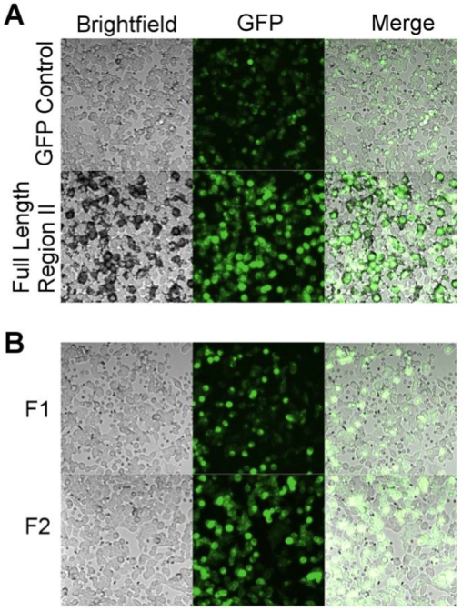
Full length RII of PfEBA-140 binds extensively to erythrocytes (top image), while individual DBL domains show little binding. This indicates that both domains are necessary for erythrocyte binding.
Licensing
{{subst:Non-commercial from license selector}}
File history
Click on a date/time to view the file as it appeared at that time.
| Date/Time | User | Dimensions | File size | Comment | |
|---|---|---|---|---|---|
| (current) | 18:49, 27 February 2023 | Sloan August (Talk | contribs) | 640×434 | 63 KB | |
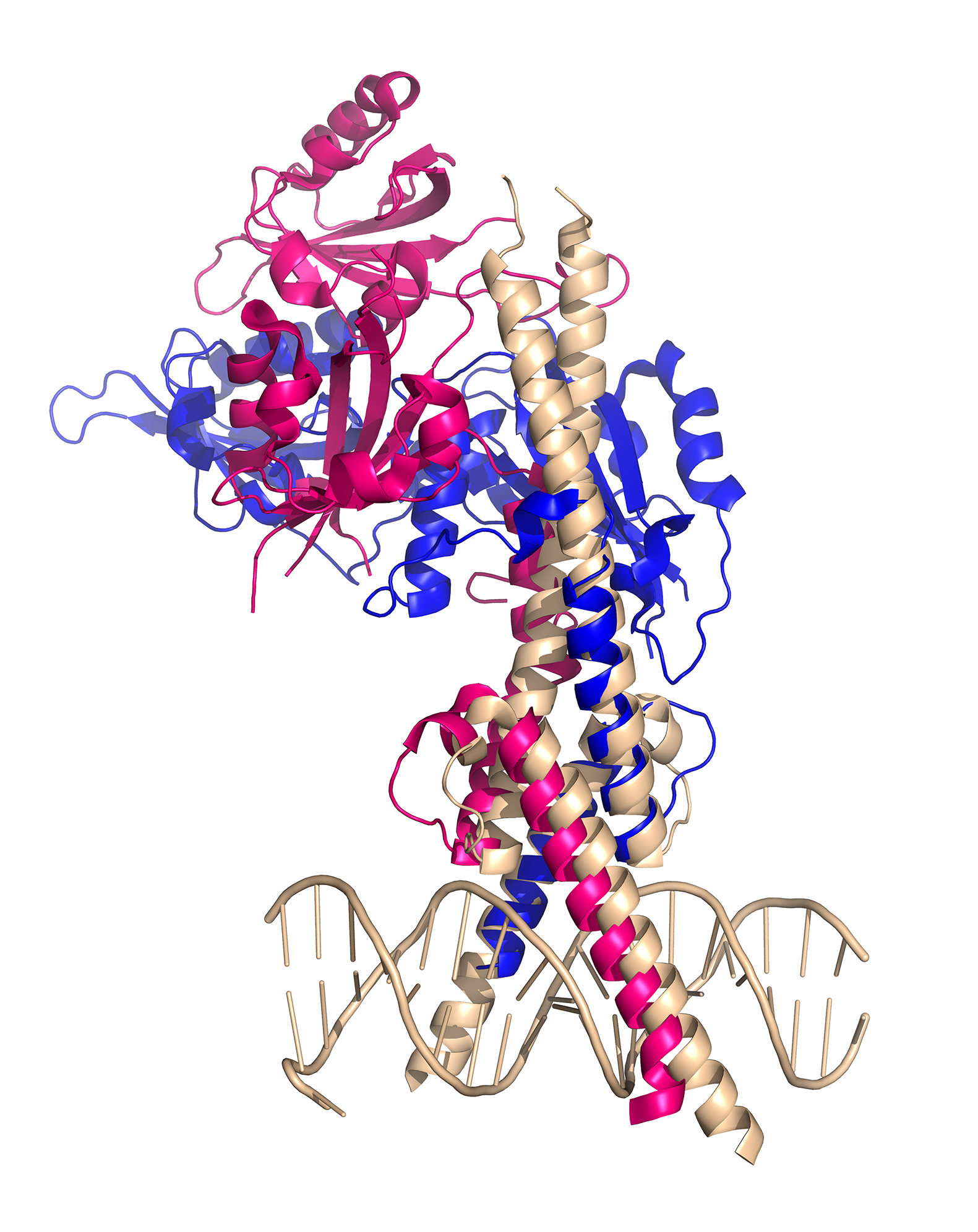
| 00:13, 3 December 2014 | Hui-Hsien Lin (Talk | contribs) | 1581×1992 | 1.24 MB | CLOCK:BMAL1 complex binds to E-box element | |
| 09:36, 14 November 2012 | Emily Lum (Talk | contribs) | 508×672 | 493 KB | Full length RII of PfEBA-140 binds extensively to erythrocytes (top image), while individual DBL domains show little binding. This indicates that both domains are necessary for erythrocyte binding. |
- Edit this file using an external application
See the setup instructions for more information.
Links
The following pages link to this file:
