Image:Binding.png
From Proteopedia
No higher resolution available.
Binding.png (640 × 434 pixel, file size: 63 KB, MIME type: image/png)
Summary
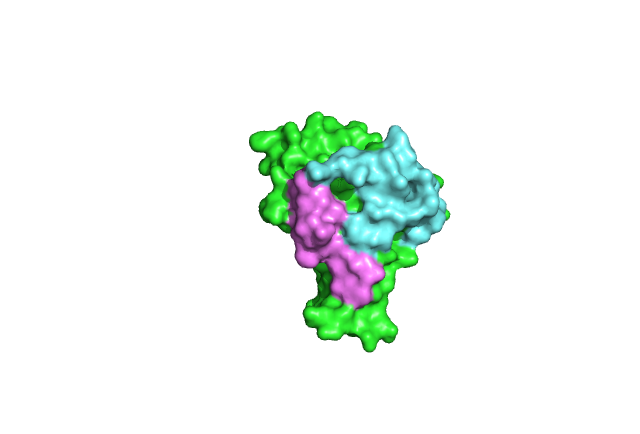
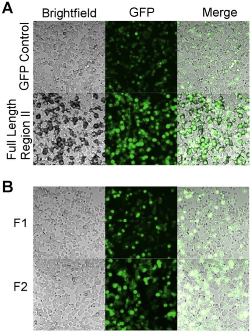
Full length RII of PfEBA-140 binds extensively to erythrocytes (top image), while individual DBL domains show little binding. This indicates that both domains are necessary for erythrocyte binding.[1]
Licensing
{{subst:Non-commercial from license selector}}
References
- ↑ Lin DH, Malpede BM, Batchelor JD, Tolia NH. Crystal and Solution Structures of Plasmodium falciparum Erythrocyte-binding Antigen 140 Reveal Determinants of Receptor Specificity during Erythrocyte Invasion. J Biol Chem. 2012 Oct 26;287(44):36830-6. doi: 10.1074/jbc.M112.409276. Epub 2012, Sep 18. PMID:22989878 doi:10.1074/jbc.M112.409276
File history
Click on a date/time to view the file as it appeared at that time.
| Date/Time | User | Dimensions | File size | Comment | |
|---|---|---|---|---|---|
| (current) | 18:49, 27 February 2023 | Sloan August (Talk | contribs) | 640×434 | 63 KB | |
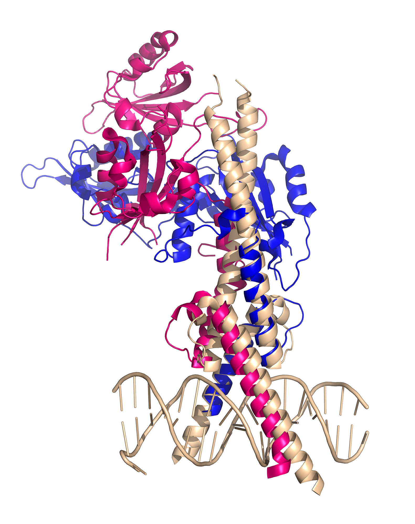
| 00:13, 3 December 2014 | Hui-Hsien Lin (Talk | contribs) | 1581×1992 | 1.24 MB | CLOCK:BMAL1 complex binds to E-box element | |
| 09:36, 14 November 2012 | Emily Lum (Talk | contribs) | 508×672 | 493 KB | Full length RII of PfEBA-140 binds extensively to erythrocytes (top image), while individual DBL domains show little binding. This indicates that both domains are necessary for erythrocyte binding. |
- Edit this file using an external application
See the setup instructions for more information.
Links
The following pages link to this file:
