Image:CuII coordination complex.jpg
From Proteopedia
No higher resolution available.
CuII_coordination_complex.jpg (387 × 290 pixel, file size: 28 KB, MIME type: image/jpeg)
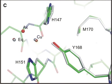
Three-dimensional structure of CuBD [1]
This figure was made thanks to pyMol program. We can see there the Cu(I) coordination geometry in CuBD, (PDB id: [http://www.rcsb.org/pdb/explore.do?structureId=2fk2 2fk2 Four aminoacids are forming the square pyramidal (tyr 168, his 147, his 151, and met170) There is also to water molecules (in red) : one is axial, and the other is equatorial.
references
- ↑ Kong GK, Adams JJ, Harris HH, Boas JF, Curtain CC, Galatis D, Masters CL, Barnham KJ, McKinstry WJ, Cappai R, Parker MW. Structural studies of the Alzheimer's amyloid precursor protein copper-binding domain reveal how it binds copper ions. J Mol Biol. 2007 Mar 16;367(1):148-61. Epub 2006 Dec 21. PMID:17239395 doi:10.1016/j.jmb.2006.12.041
File history
Click on a date/time to view the file as it appeared at that time.
| Date/Time | User | Dimensions | File size | Comment | |
|---|---|---|---|---|---|
| (current) | 23:42, 4 January 2013 | Andréa Mc Cann (Talk | contribs) | 387×290 | 28 KB | Three-dimensional structure of CuBD <ref>PMID : 17239395 </ref> This figure was made thanks to pyMol program. We can see there the Cu(I) coordination geometry in CuBD, (PDB id: [http://www.rcsb.org/pdb/explore.do?structureId=2fk2 2fk2 Four aminoacids ar |
- Edit this file using an external application
See the setup instructions for more information.
Links
The following pages link to this file:
