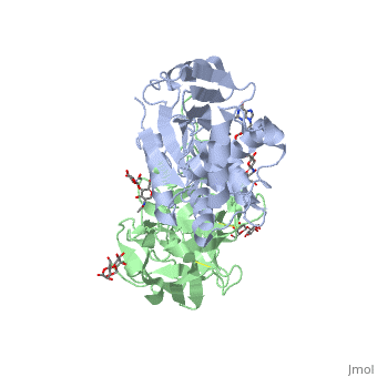
Structure
Ricin is a heterodimer that consists of a 32 kilodalton A chain glycoprotein (light blue) linked by a to a 32 kilodalton glycoprotein[2] (green).
The is an alpha/beta protein which contains eight alpha helices (pink) and eight beta sheets (yellow). It has three domains[3]. consists of a beta sheet containing both parallel and anti-parallel strands. The makes up the core of the protein, and includes the active site. The interacts with the B chain, and contains a helix and two beta strands.
The A chain contains the active site that is responsible for inactivating the Ribosome via depurination. RIPs have very diverse structures, containing only eight invariant residues[1]. These are clustered in the active site.
The B chain is a lectin[1] that to galactose-containing surface receptors. Originally it was thought that the mode of action of Ricin poisoning was due to hemagglutination due to a closely related, co-isolating lectin, RCA.
Mechanism of action
The mechanism deployed by Ricin to gain entry to a host cell involves the poison's heterogenic properties. First, the B subunit binds to two carbohydrates on the cell surface, either glycolipids or glycoproteins, which both terminate with galactose. The interaction is facilitated by hydrogen bonds to in one domain[4] and in the other domain. Once bound, the ricin-glycoprotein complex is taken into the cells via endocytosis. This association between the A and B chain is essential for toxicity [2] without it the Ricin would not be able to gain access to the cell, rendering it useless[5]. The endocytotic pathway results in the cleavage of the disulfide bond linking the A and B chains. After cleavage, the A chain is released into the cytosol.
Once the A chain gains into the cytosol, it depurinates a single adenosine residue in a highly conserved portion within the large ribosomal subunit[5] of eukaryotes; in human, the large cytoplasmic ribosomal RNA is called the 28S ribosomal RNA because of its sedimentation properties during ultracentrifugation. The nucleotide depurinated is located within a specific, conserved loop referred to as the 'sarcin-ricin loop'. Depurination of the single adenosine nucleotide by the toxin results in the inhibition of protein synthesis.
The proposed mechanism of depurination utilizes the in the A chain. The aromatic ring structures of the substrate adenosine stack with the aromatic side chains of , Tyr 80 and 123, above and below. Hydrogen bonds form between the conserved arginine and a backbone carbonyl. The depurination reaction is aided by the protonation of N3 by Arg 180 and by ion pairing to Glu 177. A water molecule on the opposite side of the ribose is activated by hydrogen bonding to Arg 180. The activated water attacks C1' of the ribose, releasing the adenine and depurinated RNA fragment. This interferes with elongation factor binding to the ribosome, thus inhibiting translation.