Maureen E. Hill/Sandbox1
From Proteopedia
Caspases are a family of CBI Molecules being studied in the University of Massachusetts Amherst Chemistry-Biology Interface Program at UMass Amherst and on display at the Molecular Playground.
Executioner Caspase-7
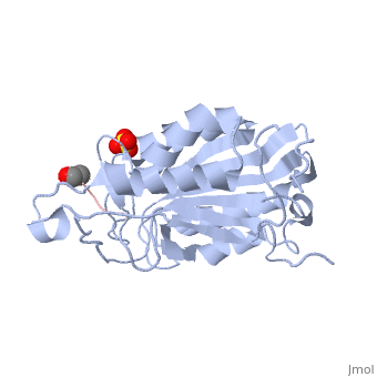
BackgroundCaspases are cysteine-aspartate proteases that are responsible for the execution of apoptosis, also known as programmed cell death. Dysregulation of apoptosis has been linked to neurodegenerative disorders, including Alzheimer's and Huntington's, as well as inflammatory diseases and cancer. The apoptotic caspases consist of two distinct classes: the initiators (caspase -2, -8, -9, and -10) and the executioners (caspase -3, -6, and -7). All caspases are synthesized as catalytically inactive zymogens that must undergo proteolytic cleavage to be activated during apoptosis. Initiator caspases are activated by upstream cellular events, which in turn cleave at distinct internal aspartate residues in the executioner caspases to remove the prodomain and separate the large and small subunits. The executioner caspases then cleave a wide range of targets within the cell that ultimately leads to cellular suicide. Caspase-7 StructureCaspases are crystallized as homodimers. As previously stated, the caspases undergo proteolytic cleavage by the initiator caspases to assume their active conformations. Some caspases undergo more cleave than others. For executioner caspase-7 there exists three major cleavage sites, D23, D198 and D207. D23 processing removes the pro domain from the large subunit, where as, D198 and D207 is the major cleavage site for processing and removal of the inter-subunit linker. Caspase-7 has one minor cleavage site also located within the inter-subunit linker at D192.
The is made up of four flexible loops which include L2, L3 and L4 from one half of the dimer that interact with L2' from the opposite half of the dimer. In the , the loops are disordered, which prevents substrate binding. Upon cleavage at the intersubunit linker, the active-site loop bundle becomes partially ordered, whereas L2' stays in the inactive, down conformation. At this point, caspase-7 may bind either substrate or allosteric inhibitors. traps the protein an active/substrate bound conformation. Substrate binding forces a conformational change moving L2' upward, this creates a foundation beneath the L2 bundle stabilizing the active complex.
Allosteric Inhibition of Caspase-7It has been shown that caspases -3 and -7 can be inhibited at a site other than the active site by allosteric inhibitors, such as 5-Fluoro-1H-indole-2-carboxylic acid (2-mercapto-ethyl)-amide (FICA) or 2-(2,4-Dichlorophenoxy)-N-(2-mercapto-ethyl)-acetamide (DICA), at the dimer-interface cavity. The mechanism of these inhibitors lock the L2' in a down conformation, thereby inactivating the enzyme. at the dimer interface. The allosteric inhibitor binds to C290 within the dimer interface displacing Y223. The displacement of tyrosine from the active site conformation of the enzyme forces R187 into a position that both physically blocks substrate binding, as well as, move the active site cystine 186. Ultimately, these conformational changes inactivate the enzyme.
Forms of Caspase-7, trapping protein in active/substrate bound conformation. trapping protein in a form incompatible with substrate binding. of Caspase-7.
ReferencesHardy, J. A., J. Lam, et al. (2004). "Discovery of an allosteric site in the caspases." Proc Natl Acad Sci U S A 101(34): 12461-12466. Witkowski, W. a, & Hardy, J. a. (2009). L2’ loop is critical for caspase-7 active site formation. Protein science : a publication of the Protein Society, 18(7), 1459–68. doi:10.1002/pro.151 Witkowski, W. a, & Hardy, J. a. (2011). A designed redox-controlled caspase. Protein science : a publication of the Protein Society, 20(8), 1421–31. doi:10.1002/pro.673 | ||||||||||||