Image:H2A.png
From Proteopedia
Size of this preview: 496 × 599 pixels
Full resolution (550 × 664 pixel, file size: 216 KB, MIME type: image/png)
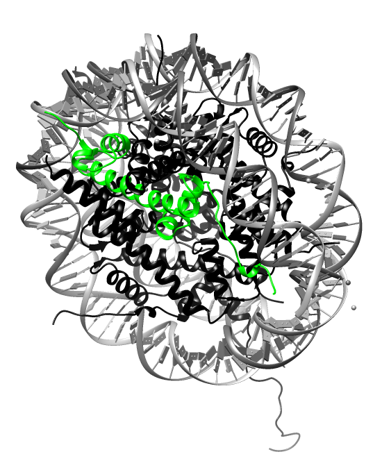
H2A protein highlighted in nucleosome model, PDB:1aoi
Molecular graphics and analyses were performed with the UCSF Chimera package. Chimera is developed by the Resource for Biocomputing, Visualization, and Informatics at the University of California, San Francisco (supported by NIGMS P41-GM103311).
File history
Click on a date/time to view the file as it appeared at that time.
| Date/Time | User | Dimensions | File size | Comment | |
|---|---|---|---|---|---|
| (current) | 17:57, 2 May 2014 | A. Rahim Zalal (Talk | contribs) | 550×664 | 216 KB | H2A protein highlighted in nucleosome model PDB:1aoi Molecular graphics and analyses were performed with the UCSF Chimera package. Chimera is developed by the Resource for Biocomputing, Visualization, and Informatics at the University of California, San |
- Edit this file using an external application
See the setup instructions for more information.
Links
The following pages link to this file:
