Publication Abstract from PubMed
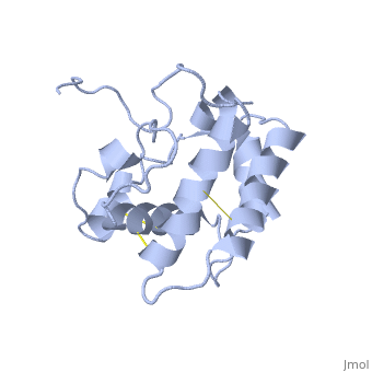
The nuclear magnetic resonance structure of the unliganded pheromone-binding protein (PBP) from Bombyx mori at pH above 6.5, BmPBP(B), consists of seven helices with residues 3-8, 16-22, 29-32, 46-59, 70-79, 84-100, and 107-124, and contains the three disulfide bridges 19-54, 50-108, and 97-117. This polypeptide fold encloses a large hydrophobic cavity, with a sufficient volume to accommodate the natural ligand bombykol. The polypeptide folds in free BmPBP(B) and in crystals of a BmPBP-bombykol complex are nearly identical, indicating that the B-form of BmPBP in solution represents the active conformation for ligand binding.
NMR structure of the unliganded Bombyx mori pheromone-binding protein at physiological pH.,Lee D, Damberger FF, Peng G, Horst R, Guntert P, Nikonova L, Leal WS, Wuthrich K FEBS Lett. 2002 Nov 6;531(2):314-8. PMID:12417333[1]
From MEDLINE®/PubMed®, a database of the U.S. National Library of Medicine.