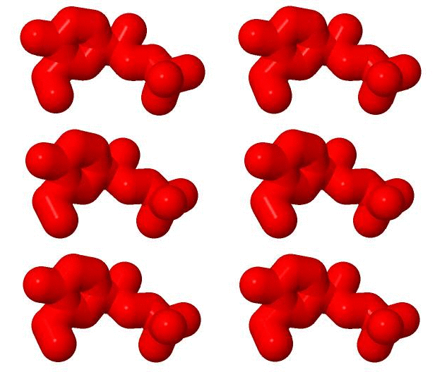
It turns out that some molecules, such as the drug salbutamol we are using as an example here, are different from their mirror image. The left image shows the actual drug, while the browser on the right shows its mirror image. If you click on the , we'll show the actual drug in the browser as well, and you can rotate it with the mouse to show how they match.
So far we focused on the shape of the molecule. To fully understand how salbutamol binds, we should show you what atoms it is made of. If you click on the "Show the atoms" link below, carbon atoms will appear in gray, oxygen atoms in red, and nitrogen atoms in blue. The molecule also contains hydrogen atoms, but these are much smaller and are not shown.
What about the box?
When salbutamol acts in the body, it does so by binding to a receptor in the cell membrane. Receptors help to communicate between the outside of the cell and the inside of the cell, much like a window or a door bell helps people in a house learn about what is going on outside. The receptor salbutamol binds to is a protein called the
It's a big molecule, and salbutamol is difficult to see because it is surrounded by the receptor. Use the links below to "shave away" parts of the molecule for a better look inside
The next scene shows salbutamol in red, and those parts of the receptor that surround it, called its binding site, in blue. Compare the blue parts to the box in the movie. It helps to use the "shave away" buttons above, and to rotate the view so it matches the view of the molecule in the movie.
Leftovers
.
Function
Disease
Relevance
Structural highlights
This is a sample scene created with SAT to by Group, and another to make of the protein. You can make your own scenes on SAT starting from scratch or loading and editing one of these sample scenes.

