User:Rana Saad/The human GABAb receptor
From Proteopedia
|
This is a default text for your page Human gabab receptor. Click above on edit this page to modify. Be careful with the < and > signs. You may include any references to papers as in: the use of JSmol in Proteopedia [1] or to the article describing Jmol [2] to the rescue. ---
Contents |
Introduction
γ-Aminobutyric acid (GABA)
GABA is the major inhibitory neurotransmitter in the central nervous system (CNS). It plays a key role in modulating neuronal activity since it binds to specific transmembrane receptors (GABAa,GABAb and GABAc) in the plasma membrane of both pre- and postsynaptic neuronal level.
GABAb receptors
Mammalian GABAb receptor is a class C G-protein coupled receptor[3]. Its structure is similar to mGluR ligand binding domain. GABAb is central to inhibitory neurotransmission in the brain and so is considered a good candidate for treatments against alcoholism, stress and number of brain diseases[4].
Function
The GABAb receptor causes the opening of the K+ channels in the postsynaptic membrane, bringing the neuron closer to the equilibrium potential of K+, producing hyperpolarization. As a result the Ca+2 channels in the presynaptic terminal close and neurotransmitter release stops. GABAb can also reduce the activity of adenylyl cyclase and decrease the cell’s conductance to Ca+2
structure
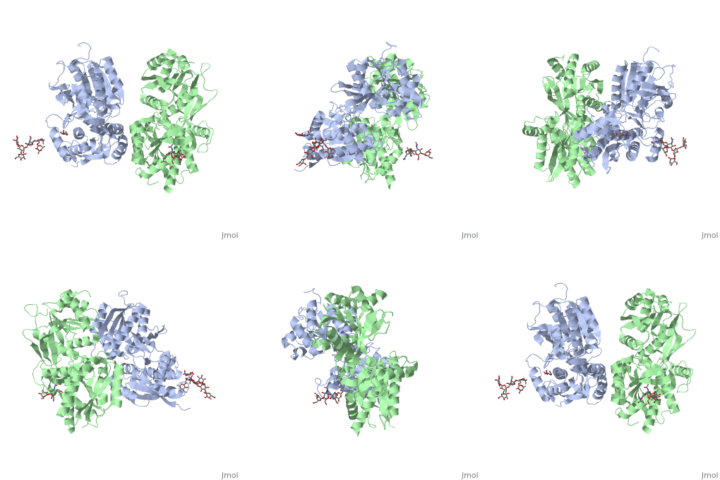
GABAb functions as an obligatory heterodimer subunit of GBR1, which is responsible for ligand-binding. GBR2, on the other hand, is responsible for G protein coupling subunits.
GBR1 and GBR2 subunits structure
Each subunit is a domain of seven-transmembrane helixes, composed of a large extracellular domain - venus flytrap (VFT). VFT contains two lobe-shaped domains: LB1 and LB2, which are connected by three short loops. LB1 and LB2 are αβ-folds composed of central β sheet flanked by an α helix
VFT interaction
Disease
Relevance
Structural highlights
This is a sample scene created with SAT to by Group, and another to make of the protein. You can make your own scenes on SAT starting from scratch or loading and editing one of these sample scenes.
</StructureSection>
References
- ↑ Hanson, R. M., Prilusky, J., Renjian, Z., Nakane, T. and Sussman, J. L. (2013), JSmol and the Next-Generation Web-Based Representation of 3D Molecular Structure as Applied to Proteopedia. Isr. J. Chem., 53:207-216. doi:http://dx.doi.org/10.1002/ijch.201300024
- ↑ Herraez A. Biomolecules in the computer: Jmol to the rescue. Biochem Mol Biol Educ. 2006 Jul;34(4):255-61. doi: 10.1002/bmb.2006.494034042644. PMID:21638687 doi:10.1002/bmb.2006.494034042644
- ↑ Stevens RC, Cherezov V, Katritch V, Abagyan R, Kuhn P, Rosen H, Wuthrich K. The GPCR Network: a large-scale collaboration to determine human GPCR structure and function. Nat Rev Drug Discov. 2013 Jan;12(1):25-34. doi: 10.1038/nrd3859. Epub 2012 Dec, 14. PMID:23237917 doi:http://dx.doi.org/10.1038/nrd3859
- ↑ Addolorato G, Leggio L, Cardone S, Ferrulli A, Gasbarrini G. Role of the GABA(B) receptor system in alcoholism and stress: focus on clinical studies and treatment perspectives. Alcohol. 2009 Nov;43(7):559-63. doi: 10.1016/j.alcohol.2009.09.031. PMID:19913201 doi:http://dx.doi.org/10.1016/j.alcohol.2009.09.031

