Sandbox Reserved 1128
From Proteopedia
Thioredoxin Reductase 1 (Human)
|
Thioredoxin reductase 1(TrxR1) is an ubiquitous enzyme which reduces the thioredoxin protein by a disulfide oxidoreductase activity [1]. This enzyme belongs to the flavoprotein family which needs cofactors to catalyze the NADPH-dependent reaction. NADPH cofactor allows electrons transmission during the reaction via FAD from enzyme to oxidized protein. The thioredoxin system is thus composed of thioredoxin reductase, NADPH and thioredoxin. The reaction catalized is:
TrxR1 belongs to one of the two forms of mammalian TrxR enzymes mostly present in cytosol contrary to TrxR2 which is only mitochondrial. TrxR1 is a heterogeneous protein which is present in most tissues and is specific of the small thioredoxin 1 protein (Trx1) [2]. Its capacity to reduce oxidized Trx1 is important to maintain the active site of Trx1. The redox activity of reduced Trx1 is the key of its biological activity [3]. TrxR1 can thus regulate Trx1 activities by its NADPH dependent reduction specificity.
Contents |
Structure
The structure is really important to understand redox regulatory processes of the human Thioredoxin reductase 1. It could be also the template for drug design because we know that this protein is involved in various diseases.
TrxR1 is a flavoprotein belonging to pyridine nucleotide-disulfide oxido-reductase protein family which is a homopolymeric protein. The functional enzyme uses thus the flavin adenine dinucleotide (FAD) as nucleic acid cofactor. TrxR is currently the only enzyme know to reduce Trx oxidized through NADPH as the source of reducing equivalents. Function of this enzyme is correlated with structural researches.
Indeed, it has been well characterized that each monomer contains thus a FAD prosthetic group, an NADPH binding site and an active site containing a redox active disulphide which acts as proton acceptor. The active site of the human TrxR1 has been also identified as a seleneylsulfide and this selenolthiol motif is formed from a sequence conserved: Cys-Val-Asn-Val-Gly-Cys [4]. Trx can interact with many partners in different cellular compartments. Its biological function is cellular localisation-dependent. Reductase activity of Trx1 can regulate cell growth or apoptosis for example, but in the nucleus, Trx can bind to different transcription factors. The key role of Trx is its capacity to defence against oxidative damage.
A central role for oxidative stress
Oxidative stress is the imbalance between oxidative and reducing species and several mediators can alternate this redox potential. Most of the time, pro-oxidative species derived from di-oxygen (O2) or nitrogen monoxide (NO) in high concentration cause oxidative damages. Reactive oxygen species (ROS) have signaling functions and their increased levels are stimuli for cells. In response, cells engage into a program to change their characteristics, such as differentiation or apoptosis.
Reductase activity of Trx, activated by TrxR1, can neutralize the ROS in order to equilibrate the oxidative drift by mediating the reduction of proteins involved in scavenging ROS. [5]. Trx1 is an important hydrogen donor to ribonucleotide reductase which has an intracellular antioxidant activity. Trx acts thus as an important regulator of the oxidative stress.
Regulation of signal transduction pathways
Some cellular pathways are affected by increase of the levels of oxidizing species below those inducing damage. Several of these pathways rely on transcriptional responses by activation of redox-sensitive transcription factors such as p53, AP-1 or NF-κB in cytoplasm [6] [7]. These transcription factors are then activated by Trx in nucleus. This enzyme over-expressed can thus bind redox-sensitive transcription factors and activates them. That leads to modulate their DNA-binding activity on the promoter region of several genes. Transcription factors regulate in this way expression of genes which leads to cellular activation and regulates apoptosis [8]. For example, the tumour suppressor protein p53 stimulates reporter gene expression involved in cellular function such as mitosis or apoptosis. He is the guardian of the genome in prevent mutations by inducing expression of various genes as redox related genes, apoptosis related genes and many other [9]. In addition, Trx can also regulate the transcription factor NF-κB which is involved in the control of several processes as cell growth, immune response or even inflammation [10].
Impact on the immune system
Thioredoxin was first identified as a cytokine like factor in virus transformed cells [11]. Indeed, the Trx protein allows reduce NF-κB by its binding to this transcription factor. NF-κB factor can be thus a redox sensitive factor by regulation of gene expression of cytokines or other immune response genes. Trx allows directly regulation of pro-inflammatory cytokines expression and demonstrates several anti-inflammatory effects [12].
Diseases
Cancer
Studies showed that Thioredoxine reducatase 1 (TrxR1) is overexpressed (10 times than the normal)[13]. in many cancer cells. Even if the direct link between TrxR1 and cancer induction is not clear, there is evidence that TrxR1 regulates DNA replication[14]. Reduction in TrxR1 activity results in blockage of cells in phase S. Indeed, decrease in TrxR1 concentration leads to decrease in reduced-Trx (activated form) which normally acts as an electron donor for the Ribonucleotide reductase (the enzyme that syntehises most deoxyribonucleotides from ribonucleotides). Some TrxR1 isoforms are thought to guide actin and tubulin polymerization[15]. Inactive-form Trx are also unable to inhibit apoptosis, as unable to inhibit the Apoptosis Signal-regulating Kinase 1 (ASK1), also known as Mitogen-Activated Protein Kinase Kinase Kinase 5 (MAP3K5). Furthermore, a studie showed that the Thioredoxin system, cooperating with the redox factor-1 (APE/Ref-1) regulates basal activity of p53 [16]. TrxR1 is then a good target for cancer treatement. Recent studies showed that arsenic trioxide AS2O3 or ATO inhibits TrxR1[17] in its redox state in presence of NADPH and is an approved anti-cancer chemotherapeutic drug commercially named Trisenox®[18]. It is used to treat acute promyelocytic leukemia, multiple myeloma, chronic myelogenous leukemia, and acute myelogenous leukemia.

Cardiovascular diseases
Thioredoxin Reductase 1 (TrxR1) has important protection functions against oxidative stress due to its antioxidant properties. Studies showed that downregulation of TrxR1 inducing Trx downregulation leads to cardiomyocyte injuries. TrxR, indirectly via TrX, plays a protective role in myocardial ischemia/reperfusion[19]. TrxR1, part of the Thioredoxin system is suggested to play a role in cellular defense against oxidized LDL and arteriosclerosis development. TrxR could also be involved in hypertension and diabetes as anormal levels of thioredoxin is found in hypertensive and diabetic patients serum compared to normal patients[20].
Neuronal diseases
Thioredoxin Reductase 1 (TrxR1) could be related to Parkinston’s Disease as TrxR1 expression decreased in the substantia nigra pars compacta of the Parkinson's disease mouse model, suggesting that TrxR1 may play a protective antioxidant role preventing neurodegeneration[21]. Although the direct involvement of Thioredoxin Reductase 1 is not clear in Alzheimer disease, studies showed that patients diagnosed with Alzheimer have a higher level of Thioredoxin Reductase 1 (TrxR1) in their brain[22].
AIDS and inflamatory diseases
Thioredoxin Reductase 1 (TrxR1) plays an important role in repressing HIV-1 protease by negatively regulating the HIV-1 transcriptional activator in human macrophages[23]. Studies suggested that TrxR1, by regulating ROS generation, regulate the redox-dependent signal transduction and may confer protection against inflammatory processes induced by influenza virus[24].
</StructureSection>
References
- ↑ Mustacich D, Powis G. Thioredoxin reductase. Biochem J. 2000 Feb 15;346 Pt 1:1-8.
- ↑ Jurado J, Prieto-Alamo MJ, Madrid-Rísquez J, Pueyo C. Absolute gene expression patterns of thioredoxin and glutaredoxin redox systems in mouse. J Biol Chem. 2003 Nov 14;278(46):45546-54. Epub 2003 Sep 3.
- ↑ Holmgren A, Björnstedt M. Thioredoxin and thioredoxin reductase. Methods Enzymol. 1995;252:199-208.
- ↑ Lothrop AP, Snider GW, Ruggles EL, Patel AS, Lees WJ, Hondal RJ. Selenium as an electron acceptor during the catalytic mechanism of thioredoxin reductase. Biochemistry. 2014 Feb 4;53(4):654-63. doi: 10.1021/bi400658g. Epub 2014 Jan 23.<\ref>.
This protein has a high molecular weight: each monomer has a molecular mass of 54,6 kDa.
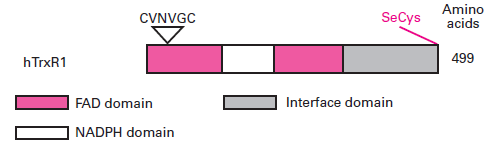 The results reported by Sandalova and tal. (2001) showed the crystal structure analysis of the SeCys498Cys mutant of rat TrxR complexed with NADP+ <ref>Sandalova T, Zhong L, Lindqvist Y, Holmgren A, Schneider G. Three-dimensional structure of a mammalian thioredoxin reductase: implications for mechanism and evolution of a selenocysteine-dependent enzyme. Proc Natl Acad Sci U S A. 2001 Aug 14;98(17):9533-8. Epub 2001 Jul 31.<\ref>. That allowed visualizing the architecture of the active site in an overall fold of the enzyme.
[[Image:Structure.png]]
Each binding domain have a central five-stranded parallel β-sheet and three-stranded β-meander. In the other side of parallel sheet there are several α-helices. The two cysteins Cys59 and Cys64 which form the active disulfide, are located on helix α2. The interaction of the two subunits takes place at the interface domain which contains antiparallele five-stranded β-sheet E flanked on both sides by four helices. The C terminal domain contains an extension with a selenocysteine (Se) residue with specific motif of Gly-Cys-SeCys-Gly. The first three residues of the extension continue in the direction toward the surface of the molecule.
== Catalytic mechanism ==
Se-containing TrxR has a catalytic action on the reduction reaction of Trx.
The FAD domain which contain the active site (catalytic site) and the binding site of NADPH are close to the ring of FAD. That allows the electrons to go from NADPH to the substrate through the ring of FAD and the disulphide active site. There is no big conformational change in the enzyme to do that. When there is an excess of NADPH, hTrxR forms a stable charge transfer complex.
In hTrxR there is an additional redox-active site which is not part of the conserved active site.
The action mechanism of Se-containing TrxR is supposed to be like that:
The results reported by Sandalova and tal. (2001) showed the crystal structure analysis of the SeCys498Cys mutant of rat TrxR complexed with NADP+ <ref>Sandalova T, Zhong L, Lindqvist Y, Holmgren A, Schneider G. Three-dimensional structure of a mammalian thioredoxin reductase: implications for mechanism and evolution of a selenocysteine-dependent enzyme. Proc Natl Acad Sci U S A. 2001 Aug 14;98(17):9533-8. Epub 2001 Jul 31.<\ref>. That allowed visualizing the architecture of the active site in an overall fold of the enzyme.
[[Image:Structure.png]]
Each binding domain have a central five-stranded parallel β-sheet and three-stranded β-meander. In the other side of parallel sheet there are several α-helices. The two cysteins Cys59 and Cys64 which form the active disulfide, are located on helix α2. The interaction of the two subunits takes place at the interface domain which contains antiparallele five-stranded β-sheet E flanked on both sides by four helices. The C terminal domain contains an extension with a selenocysteine (Se) residue with specific motif of Gly-Cys-SeCys-Gly. The first three residues of the extension continue in the direction toward the surface of the molecule.
== Catalytic mechanism ==
Se-containing TrxR has a catalytic action on the reduction reaction of Trx.
The FAD domain which contain the active site (catalytic site) and the binding site of NADPH are close to the ring of FAD. That allows the electrons to go from NADPH to the substrate through the ring of FAD and the disulphide active site. There is no big conformational change in the enzyme to do that. When there is an excess of NADPH, hTrxR forms a stable charge transfer complex.
In hTrxR there is an additional redox-active site which is not part of the conserved active site.
The action mechanism of Se-containing TrxR is supposed to be like that:
- • The NADPH fixes to the TrxR NADPH binding site and the electrons go from NADPH to the conserved catalytic site through FAD to reduce the disulphide bond of TrxR. So at this step the catalytic site is in its oxidized form.
- • Then there is the oxidation of the Cys-X-X-Cys site and the reduction of the C-terminal Se-containing site thanks to a Thiol-Disulfide exchange.
- • The reduced C-terminal site goes away transferring electrons to a substrate.

