Sandbox Reserved 430
From Proteopedia
| This Sandbox is Reserved from January 19, 2016, through August 31, 2016 for use for Proteopedia Team Projects by the class Chemistry 423 Biochemistry for Chemists taught by Lynmarie K Thompson at University of Massachusetts Amherst, USA. This reservation includes Sandbox Reserved 425 through Sandbox Reserved 439. |
P2Y12 Receptor in Complex with AZD1283 (4ntj)[1]
by [Cora Ricker, Lauren Timmins, Aidan Finnerty, Adam Murphy, Duy Nguyen]
Student Projects for UMass Chemistry 423 Spring 2016
IntroductionThe goal of pharmaceuticals is to prevent or cure disease through drug therapy by specifically targeting cells, proteins, enzymes, genes, etc. It is often crucial to understand the structure, function, and relevant mechanisms involved with the target when designing an effective drug candidate. Furthermore, being able to know the effects on structure after drug-binding can provide insight into the functionality of a specific target. Through crystal structure analysis of P2Y12 binded to AZD1283, differences in secondary and tertiary structure can be observed with and without the small molecule. This structural modifications support proposed mechanisms of active P2Y12 and provide a basis for future drug candidates. P2Y12 is a G-protein-coupled receptor (GPCR) found on blood platelets. As a GPCR, P2Y12 responds to concentrations of adenosine diphosphate in the extracellular matrix. This leads to purinergic signal transduction and ultimately cellular response. Being located on blood platelets, activated P2Y12 receptors induce platelet activation and clotting. Drugs, such as clopidogrel, ticagrelor, and ticlopidine, are designed to inhibit P2Y12 activation by altering the receptor active site through secondary and tertiary structure modification. Successful inhibition of P2Y12 corresponds to decreased platelet activation, a helpful tool in prevention of stroke and heart attacks. Additionally, P2Y12-specific drugs can be used in treating cardiovascular disease. Here, image analysis of P2Y12 crystals are used to model protein structure in complex with AstraZeneca’s novel ADZ1283: Ethyl 6-(4(-((benzylsulphonyl)carbamoyl)piperidin-1yl)-5-cyano-2-methylnicotinate. ADZ1283 functions to block the P2Y12 receptor as a means to treat thrombosis. ADZ1283-binding leads to unique protein structure, unfound in other P2Y receptors. Helix V of seven transmembrane helices is found to be . This change along with the discovery of a potential second active within P2Y12 has implications on how P2Y12 uses it’s seven transmembrane helical bundle interact with ADP in the bloodstream. a view of the Anionic Side Chains in red and charged nucleic acids and ligands in grey. Overall StructureThe is made up of eight alpha helices. There are seven transmembrane alpha helices and a carboxy-terminal helix VII, which is parallel to the membrane bilayer. There is only one disulphide bond the amino terminus with helix VII Helix V: straight conformation because there are no proline or glycine residues to destabilize its structure
- Polar vs nonpolar: Polar regions are pink. Nonpolar, or hydrophobic, regions are grey. -N to C termini: The protein shown in displays the the N and C termini
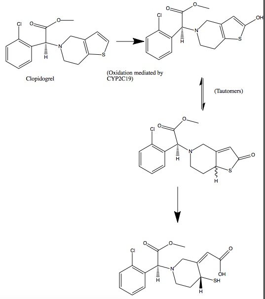
Binding InteractionsThe of P2Y12 is colored in purple, and the ligand (AZD1283) is colored in yellow. The binding site for AZD1283, pocket 1, is composed of helices III–VII, while pocket II contains helices I-III and VII is not represented here. The two pockets are separated by two residues, Y1053 and K2807, which makes the pocket II not a binding site for AZD1283. With the observation of the AZD1283-bound P2Y12R with residue C97 belong to pocket 2, the active metabolites drugs might occupy this pocket. Additional FeaturesAcute coronary syndrome, a condition in which there is sudden blockage of blood flow to the heart, is majorly caused by a disease known as atherothrombosis. This disease is characterized by blocked arteries due to thrombosis: formation of a clot within a blood vessel. The P2Y12 protein is an important platelet receptor inhibitor that can be combined with aspirin in the management of ACS. It regulates certain functions through purinergic signaling, which is a form of extracellular signaling mediated by purine nucleotides and nucleosides like ADP and ATP. The P2Y12 receptor is mainly found on the surface of blood platelets, and functions as a regulator in platelet activation and therefore blood clotting. The P2Y12 receptor is a G-protein coupled receptor, linked to the cAMP-signaling pathway. It mediates platelet activation by decreasing intracellular cAMP levels through inhibition of an AC-mediated signaling pathway. Acting as a chemoreceptor, P2Y12 utilizes ADP as an agonist, which initiates ADP-induced platelet aggregation. Clopidogrel in covalent binding complex with P2Y12 acts as an antiplatelet agent, specifically as an ADP receptor inhibitor to decrease platelet aggregation and inhibit thrombus formation. Clopidogrel is a pro-drug and thienopyridine-type inhibitor of the P2Y12 receptor, which requires Cytochrome P450 to hepatically transform it to exert its anti platelet effect. is a membrane protein, characteristic of alternating hydrophobic and polar groups. The central heme group is stabilized by several side chains. The atom that makes up the heart of the heme group is surrounded by a highly hydrophobic porphyrin ring . The role of the heme group in biological systems is to facilitate oxygen transport as well as aid in electron transfer as part of the electron transport chain. The heme group of the hemeprotein Cytochrome P450 acts as a catalyst for the metabolism of clopidogrel. Shown below is the activation of Clopidogrel in vivo. CYP2C19 is an enzymatic member of the cytochrome P450 mixed-function oxidase system.
Quiz Question 1What other residues can be used to replace the ? See AlsoCreditsIntroduction - Adam Murphy Overall Structure - Cora Ricker Drug Binding Site - Duy Nguyen Additional Features - Lauren Timmins Quiz Question 1 - Aidan Finnerty References
| ||||||||||||

