This old version of Proteopedia is provided for student assignments while the new version is undergoing repairs. Content and edits done in this old version of Proteopedia after March 1, 2026 will eventually be lost when it is retired in about June of 2026.
Apply for new accounts at the new Proteopedia. Your logins will work in both the old and new versions.
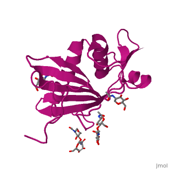
GP1 of Lassa Virus
From Proteopedia
| |||||||||||
References
- ↑ Maiztegui JI, McKee KT Jr, Barrera Oro JG, Harrison LH, Gibbs PH, Feuillade MR, Enria DA, Briggiler AM, Levis SC, Ambrosio AM, Halsey NA, Peters CJ. Protective efficacy of a live attenuated vaccine against Argentine hemorrhagic fever. AHF Study Group. J Infect Dis. 1998 Feb;177(2):277-83. PMID:9466512
- ↑ Radoshitzky SR, Abraham J, Spiropoulou CF, Kuhn JH, Nguyen D, Li W, Nagel J, Schmidt PJ, Nunberg JH, Andrews NC, Farzan M, Choe H. Transferrin receptor 1 is a cellular receptor for New World haemorrhagic fever arenaviruses. Nature. 2007 Mar 1;446(7131):92-6. Epub 2007 Feb 7. PMID:17287727 doi:http://dx.doi.org/10.1038/nature05539
- ↑ Zong M, Fofana I, Choe H. Human and host species transferrin receptor 1 use by North American arenaviruses. J Virol. 2014 Aug;88(16):9418-28. doi: 10.1128/JVI.01112-14. Epub 2014 Jun 11. PMID:24920811 doi:http://dx.doi.org/10.1128/JVI.01112-14
- ↑ Cao W, Henry MD, Borrow P, Yamada H, Elder JH, Ravkov EV, Nichol ST, Compans RW, Campbell KP, Oldstone MB. Identification of alpha-dystroglycan as a receptor for lymphocytic choriomeningitis virus and Lassa fever virus. Science. 1998 Dec 11;282(5396):2079-81. PMID:9851928
- ↑ Kunz S, Rojek JM, Perez M, Spiropoulou CF, Oldstone MB. Characterization of the interaction of lassa fever virus with its cellular receptor alpha-dystroglycan. J Virol. 2005 May;79(10):5979-87. PMID:15857984 doi:http://dx.doi.org/10.1128/JVI.79.10.5979-5987.2005
- ↑ Spiropoulou CF, Kunz S, Rollin PE, Campbell KP, Oldstone MB. New World arenavirus clade C, but not clade A and B viruses, utilizes alpha-dystroglycan as its major receptor. J Virol. 2002 May;76(10):5140-6. PMID:11967329
- ↑ Eschli B, Quirin K, Wepf A, Weber J, Zinkernagel R, Hengartner H. Identification of an N-terminal trimeric coiled-coil core within arenavirus glycoprotein 2 permits assignment to class I viral fusion proteins. J Virol. 2006 Jun;80(12):5897-907. PMID:16731928 doi:http://dx.doi.org/10.1128/JVI.00008-06
- ↑ Pasquato A, Burri DJ, Traba EG, Hanna-El-Daher L, Seidah NG, Kunz S. Arenavirus envelope glycoproteins mimic autoprocessing sites of the cellular proprotein convertase subtilisin kexin isozyme-1/site-1 protease. Virology. 2011 Aug 15;417(1):18-26. doi: 10.1016/j.virol.2011.04.021. Epub 2011, May 25. PMID:21612810 doi:http://dx.doi.org/10.1016/j.virol.2011.04.021
- ↑ Burri DJ, da Palma JR, Kunz S, Pasquato A. Envelope glycoprotein of arenaviruses. Viruses. 2012 Oct 17;4(10):2162-81. doi: 10.3390/v4102162. PMID:23202458 doi:http://dx.doi.org/10.3390/v4102162
- ↑ 10.0 10.1 Cohen-Dvashi H, Cohen N, Israeli H, Diskin R. Molecular mechanism for LAMP1 recognition by Lassa Virus. J Virol. 2015 May 13. pii: JVI.00651-15. PMID:25972533 doi:http://dx.doi.org/10.1128/JVI.00651-15
- ↑ Cao W, Henry MD, Borrow P, Yamada H, Elder JH, Ravkov EV, Nichol ST, Compans RW, Campbell KP, Oldstone MB. Identification of alpha-dystroglycan as a receptor for lymphocytic choriomeningitis virus and Lassa fever virus. Science. 1998 Dec 11;282(5396):2079-81. PMID:9851928
- ↑ Kunz S, Rojek JM, Perez M, Spiropoulou CF, Oldstone MB. Characterization of the interaction of lassa fever virus with its cellular receptor alpha-dystroglycan. J Virol. 2005 May;79(10):5979-87. PMID:15857984 doi:http://dx.doi.org/10.1128/JVI.79.10.5979-5987.2005
- ↑ 13.0 13.1 Eschli B, Quirin K, Wepf A, Weber J, Zinkernagel R, Hengartner H. Identification of an N-terminal trimeric coiled-coil core within arenavirus glycoprotein 2 permits assignment to class I viral fusion proteins. J Virol. 2006 Jun;80(12):5897-907. PMID:16731928 doi:http://dx.doi.org/10.1128/JVI.00008-06
- ↑ Li S, Sun Z, Pryce R, Parsy ML, Fehling SK, Schlie K, Siebert CA, Garten W, Bowden TA, Strecker T, Huiskonen JT. Acidic pH-Induced Conformations and LAMP1 Binding of the Lassa Virus Glycoprotein Spike. PLoS Pathog. 2016 Feb 5;12(2):e1005418. doi: 10.1371/journal.ppat.1005418., eCollection 2016 Feb. PMID:26849049 doi:http://dx.doi.org/10.1371/journal.ppat.1005418
- ↑ Oppliger J, Torriani G, Herrador A, Kunz S. Lassa Virus Cell Entry via Dystroglycan Involves an Unusual Pathway of Macropinocytosis. J Virol. 2016 Jun 24;90(14):6412-29. doi: 10.1128/JVI.00257-16. Print 2016 Jul, 15. PMID:27147735 doi:http://dx.doi.org/10.1128/JVI.00257-16
- ↑ 16.0 16.1 Cohen-Dvashi H, Israeli H, Shani O, Katz A, Diskin R. Role of LAMP1 Binding and pH Sensing by the Spike Complex of Lassa Virus. J Virol. 2016 Oct 28;90(22):10329-10338. Print 2016 Nov 15. PMID:27605678 doi:http://dx.doi.org/10.1128/JVI.01624-16
- ↑ Jae LT, Raaben M, Herbert AS, Kuehne AI, Wirchnianski AS, Soh TK, Stubbs SH, Janssen H, Damme M, Saftig P, Whelan SP, Dye JM, Brummelkamp TR. Virus entry. Lassa virus entry requires a trigger-induced receptor switch. Science. 2014 Jun 27;344(6191):1506-10. doi: 10.1126/science.1252480. PMID:24970085 doi:http://dx.doi.org/10.1126/science.1252480
- ↑ Israeli H, Cohen-Dvashi H, Shulman A, Shimon A, Diskin R. Mapping of the Lassa virus LAMP1 binding site reveals unique determinants not shared by other old world arenaviruses. PLoS Pathog. 2017 Apr 27;13(4):e1006337. doi: 10.1371/journal.ppat.1006337., eCollection 2017 Apr. PMID:28448640 doi:http://dx.doi.org/10.1371/journal.ppat.1006337
Categories: Cohen, N | Cohen-Dvashi, H | Diskin, R | Israeli, H | Arenavirus | Glycoprotein | Lassa | LASV | Receptor binding | Viral protein | 4zjf | GP1
