We apologize for Proteopedia being slow to respond. For the past two years, a new implementation of Proteopedia has been being built. Soon, it will replace this 18-year old system. All existing content will be moved to the new system at a date that will be announced here.
Reverse transcriptase
From Proteopedia
| |||||||||||
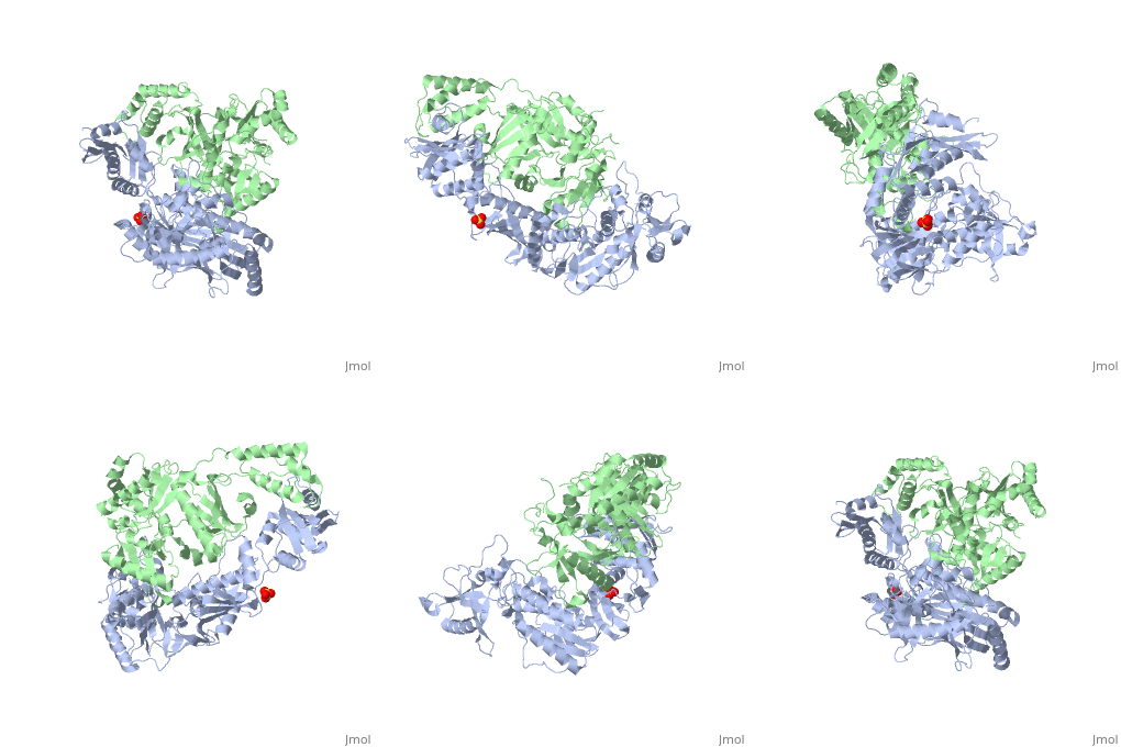
3D Structures of Reverse transcriptase
Updated on 27-September-2017
See Also
- Reverse Transcriptase at Wikipedia
- Molecule of the Month (09/2002) at RCSB Protein Data Bank
- List of Reverse Transcriptase articles at Proteopedia and at RCSB Protein Data Bank
- Model of Reverse Transcriptase as one of the CBI Molecules on the Molecular Playground
- See Transcription for additional Proteopedia articles on the subject.
- For additional information, see: Human Immunodeficiency Virus
- For additional information, see: Transcription and RNA Processing
References
- ↑ Kohlstaedt LA, Wang J, Friedman JM, Rice PA, Steitz TA. Crystal structure at 3.5 A resolution of HIV-1 reverse transcriptase complexed with an inhibitor. Science. 1992 Jun 26;256(5065):1783-90. PMID:1377403 doi:[http://dx.doi.org/10.1126/science.1377403 http://dx.doi.org/10.1126/science.1377403
- ↑ Consurf Data Base DOI: 10.1002/ijch.201200096
- ↑ DOI: 10.1002/ijch.201200096</ref Link to Consurf Data Base for PDB Entry: 1JLB. As the same rate that the polymerization process occurs, the other active site known as the cleaves RNA, releasing the ssDNA that comes again through the Polymerase active site to become dsDNA (all this with a coordinative system, that allows non-specific recognition, just with phosphates). Finally, Chain B, despite the similar aminoacid sequence with Chain A, has no enzymatic activity; its function is possibly to stabilize and interact with both active sites by varying the length between them in order to synchronize both functions. This is the most general idea of the mechanism of action of Reverse Transcriptase; however the process remains unclear and new approaches are being reported <ref> PMID: 18464735</li></ol></ref>
Proteopedia Page Contributors and Editors (what is this?)
Michal Harel, Daniel Moyano-Marino, Joel L. Sussman, Alexander Berchansky, David Canner, Amol Kapoor, Jaime Prilusky, Brian Foley, Lynmarie K Thompson, Eric Martz


