We apologize for Proteopedia being slow to respond. For the past two years, a new implementation of Proteopedia has been being built. Soon, it will replace this 18-year old system. All existing content will be moved to the new system at a date that will be announced here.
Welcome to Proteopedia ISSN 2310-6301The free, collaborative 3D-encyclopedia of proteins & other molecules
Journals
Art on Science
Selected Pages
Education
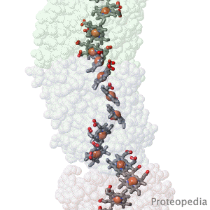
Geobacter nanowire structure surprise.
F Wang, Y Gu, JP O'Brien, SM Yi, SE Yalcin, V Srikanth, C Shen, D Vu, NL Ing, AI Hochbaum, EH Egelman, NS Malvankar. Cell 2019 doi: 10.1016/j.cell.2019.03.029 Bacteria living in anaerobic environments (no oxygen) need alternative electron acceptors in order to get energy from their food. An acceptor abundant in the earth's crust is red iron oxide ("rust"), which gets reduced to black iron oxide (magnetite). Many bacteria, such as Geobacter, get their metabolic energy by transferring electrons to acceptors that are multiple cell diameters distant, using protein nanowires. These were long thought to be pili. But when the structure of the nanowires was solved in 2019, to everyone's surprise, they turned out to be unprecedented linear polymers of multi-heme cytochromes. The hemes form an electrically conductive chain in the cores of these nanowires.
by Alice Clark (PDBe)
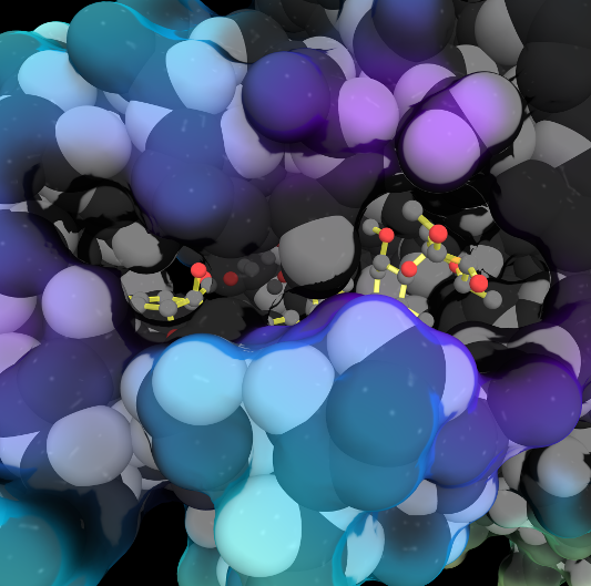
In the 1970s, an exciting discovery of a family of medicines was made by the Japanese scientist Satoshi Ōmura. One of these molecules, ivermectin, is shown in this artwork bound in the ligand binding pocket of the Farnesoid X receptor, a protein which helps regulate cholesterol in humans. This structure showed that ivermectin induced transcriptional activity of FXR and could be used to regulate metabolism.
by Eric Martz
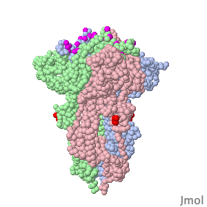
Coronavirus SARS-CoV-2 (responsible for COVID-19) has a spike protein on its surface, which enables it to infect host cells. Initially, proteases in the lungs clip the homo-trimeric spike protein at a unique sequence. This primes it, causing it to extend its receptor binding surface (shown in the above animation), optimizing binding to the host cell's ACE2 receptor (not shown). Next, spike protein initiates fusion of the virus and host cell membranes (not shown), enabling the virus RNA to enter the cell and initiate production of new virions. Knowledge of spike protein's molecular structure and function is crucial to developing effective therapies and vaccines.
>>> Visit this page >>>
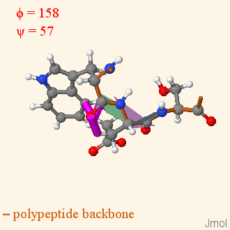
Tutorial: Ramachandran Plot Inspection
by Angel Herráez
Side-by-side display of dihedral angles in a 3D model of a tripeptide and its Ramachandran plot.
Users can interact with any of them and the other will change accordingly.
Includes animated rotations with display of clashes.