We apologize for Proteopedia being slow to respond. For the past two years, a new implementation of Proteopedia has been being built. Soon, it will replace this 18-year old system. All existing content will be moved to the new system at a date that will be announced here.
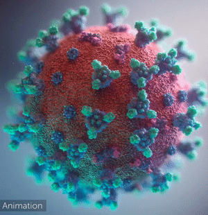
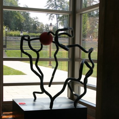
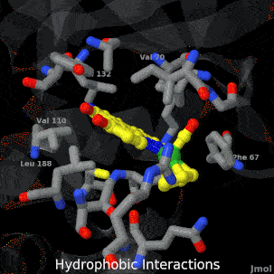
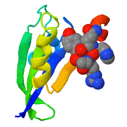
Welcome to Proteopedia ISSN 2310-6301The free, collaborative 3D-encyclopedia of proteins & other molecules
Selected Pages
Art on Science
Journals
Education
BREAKTHROUGH in protein structure prediction!
by Eric Martz
After decades of slow progress by many groups, in 2020, AlphaFold2 proved able to accurately predict the detailed structures of two-thirds of single protein domains from their amino acid sequences. Pictured is AlphaFold2's prediction for the ORF8 protein of SARS-CoV-2 (black), compared with a subsequently published X-ray crystallographic structure (colors). ORF8 contributes to virulence in COVID-19.
>>> Visit this page >>>
Opening a Gate to Human Health
by Alice Clark (PDBe)
In the 1970s, an exciting discovery of a family of medicines was made by the Japanese scientist Satoshi Ōmura. One of these molecules, ivermectin, is shown in this artwork bound in the ligand binding pocket of the Farnesoid X receptor, a protein which helps regulate cholesterol in humans. This structure showed that ivermectin induced transcriptional activity of FXR and could be used to regulate metabolism.
Structural flexibility of the periplasmic protein, FlgA, regulates flagellar P-ring assembly in Salmonella enterica.
H Matsunami, YH Yoon, VA Meshcheryakov, K Namba, FA Samatey. Scientific Reports 2016 doi: 10.1038/srep27399
A periplasmic flagellar chaperone protein, FlgA, is required for P-ring assembly in bacterial flagella of taxa such as Salmonella enterica or Escherichia coli. Here we present the open and closed crystal structures of FlgA from Salmonella enterica serovar Typhimurium, grown under different crystallization conditions. An intramolecular disulfide cross-linked form of FlgA caused a dominant negative effect on motility of the wild-type strain.
Positive (+)
and
Negative (-)
charges on the surface of a protein molecule play crucial roles in its interactions with other molecules, and hence in its functions.
Electrostatic potential maps coloring the surface of a protein molecule are a popular way to visualize the distribution
of surface charges. Easy to use free software is available to to create these surface maps.
Above is an integral membrane potassium channel protein. One of its 4 identical chains is removed so you can see the Negative (-)
protein surface contacting the
3 K+ ions.