Main Page

From Proteopedia

Revision as of 09:09, 21 October 2018 by Jaime Prilusky (Talk | contribs)
Jump to: navigation, search
ISSN 2310-6301

As life is more than 2D, Proteopedia helps to bridge the 3D relationships between function & structure of biomacromolecules


Selected Pages Art on Science Journals Education
About this image
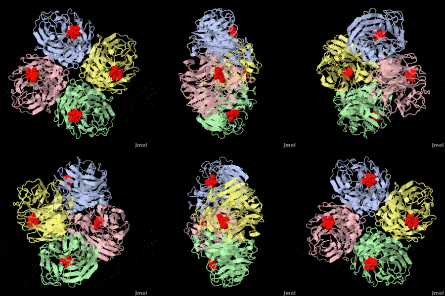
HIV-1 protease

by David Canner
The X-ray structure of HIV-1 protease reveals that it is composed of two symmetrically related subunits which form a tunnel where they meet. This is critical because it contains the active site of the protease, consisting on two Asp-Thr-Gly conserved sequences, making it a member of the aspartyl protease family. The two catalytic Asp's either interact with the incoming water or protonate the carbonyl to make the carbon more electrophilic for the incoming water.

>>> Visit this page >>>

About this image
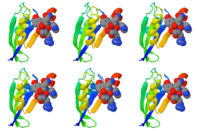
Opening a Gate to Human Health

by Alice Clark (PDBe)
In the 1970s, an exciting discovery of a family of medicines was made by the Japanese scientist Satoshi Ōmura. One of these molecules, ivermectin, is shown in this artwork bound in the ligand binding pocket of the Farnesoid X receptor, a protein which helps regulate cholesterol in humans. This structure showed that ivermectin induced transcriptional activity of FXR and could be used to regulate metabolism.

>>> Visit this page >>>

About this image
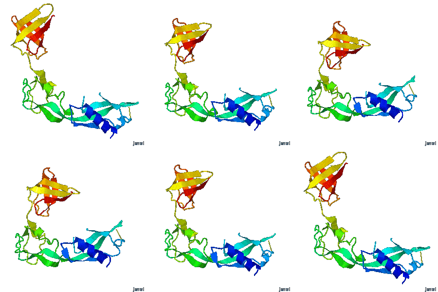
Geobacter pili: surprising function.

Y Gu, V Srikanth, AI Salazar-Morales, R Jain, JP O'Brien, SM Yi, RK Soni, FA Samatey, SE Yalcin, NS Malvankar. Nature 2021 doi: 10.1038/s41586-021-03857-w
Geobacter pili were long thought to be electrically conductive protein nanowires composed of PilA-N. Nanowires are crucial to the energy metabolism of bacteria flourishing in oxygen-deprived environments. To everyone's surprise, in 2019, the long-studied nanowires were found to be linear polymers of multi-heme cytochromes, not pili. The first cryo-EM structure of pili (2021) reveals a filament made of dimers of PilA-N and PilA-C, shown. Electrical conductivity of pili is much lower than that of cytochrome nanowires. Evidence suggests that PilA-NC filaments are periplasmic pseudopili crucial for exporting cytochrome nanowires onto the cell surface, rather than the pili serving as nanowires themselves.

>>> Visit I3DC Interactive Visualizations >>>

About this image
You Are What You Eat!

Above is an integral membrane protein that takes up, into your intestinal cells, orally consumed peptide nutrients and drugs. Its lumen-face (top) opens and binds peptide or drug (small solid object in the center), then closes, while its cytoplasmic face (bottom) opens to release its cargo into the intestinal cell, which passes it on to the blood circulation.

>>> See more animations and explanation >>>

How to add content to Proteopedia

Video Guides

Who knows ...

List of Art on Science pages in Proteopedia

What is an Interactive 3D Complement (I3DC)?

List of I3DCs

How to get an I3DC for your paper

Teaching Strategies Using Proteopedia

Examples of Pages for Teaching

How to add content to Proteopedia

About Contact Table of Contents Structure Index Help

Proteopedia Page Contributors and Editors (what is this?)

Joel L. Sussman, Jaime Prilusky

Personal tools