Main Page

From Proteopedia

Revision as of 14:19, 20 January 2019 by Joel L. Sussman (Talk | contribs)
Jump to: navigation, search
ISSN 2310-6301

As life is more than 2D, Proteopedia helps to bridge the gap between 3D structure & function of biomacromolecules

Proteopedia presents this information in a user-friendly way as a free, collaborative 3D-encyclopedia of proteins & other biomolecules.


Selected Pages Journals Education
About this image
Coronavirus Spike Protein Priming

by Eric Martz
Coronavirus SARS-CoV-2 (responsible for COVID-19) has a spike protein on its surface, which enables it to infect host cells. Initially, proteases in the lungs clip the homo-trimeric spike protein at a unique sequence. This primes it, causing it to extend its receptor binding surface (shown in the above animation), optimizing binding to the host cell's ACE2 receptor (not shown). Next, spike protein initiates fusion of the virus and host cell membranes (not shown), enabling the virus RNA to enter the cell and initiate production of new virions. Knowledge of spike protein's molecular structure and function is crucial to developing effective therapies and vaccines.
>>> Visit this page >>>

About this image
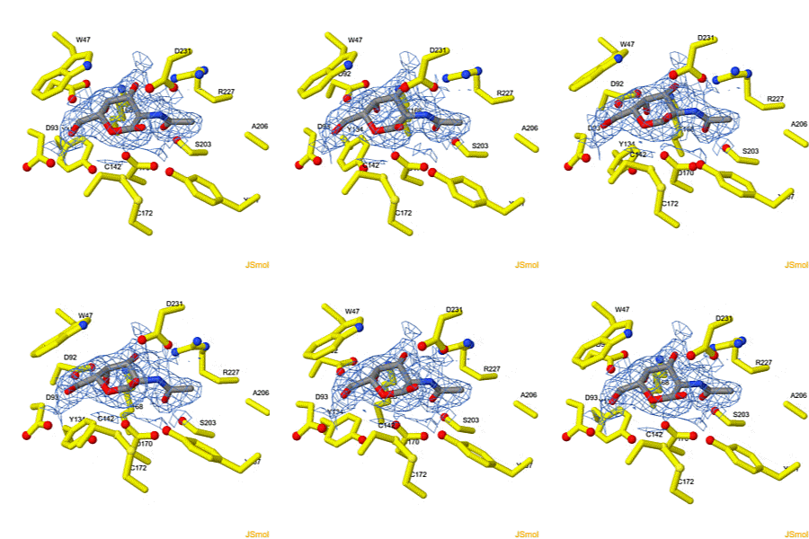
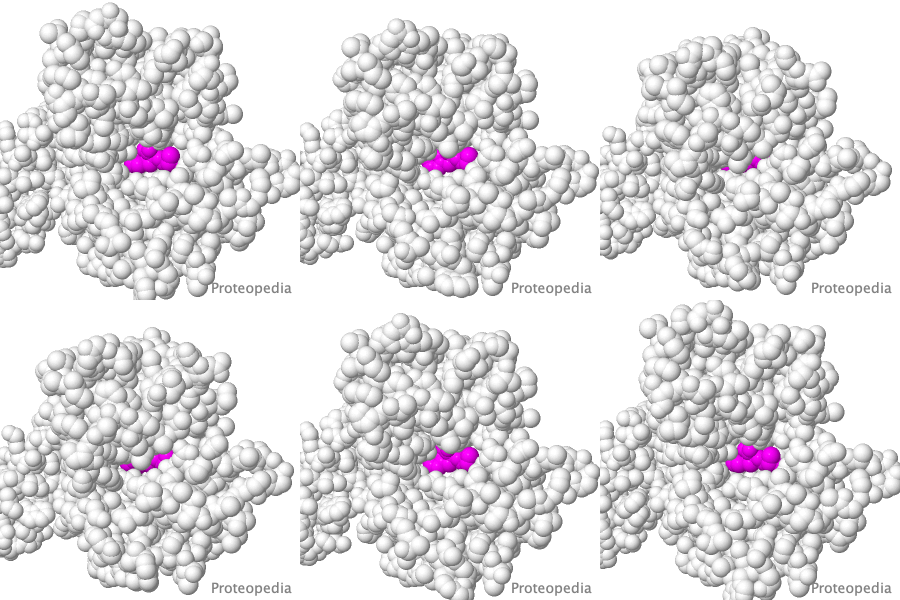
Interconversion of the specificities of human lysosomal enzymes associated with Fabry and Schindler diseases.

IB Tomasic, MC Metcalf, AI Guce, NE Clark, SC Garman. J. Biol. Chem. 2010 doi: 10.1074/jbc.M110.118588
The human lysosomal enzymes α-galactosidase and α-N-acetylgalactosaminidase share 46% amino acid sequence identity and have similar folds. Using a rational protein engineering approach, we interconverted the enzymatic specificity of α-GAL and α-NAGAL. The engineered α-GAL retains the antigenicity but has acquired the enzymatic specificity of α-NAGAL. Conversely, the engineered α-NAGAL retains the antigenicity but has acquired the enzymatic specificity of the α-GAL enzyme. Comparison of the crystal structures of the designed enzyme to the wild-type enzymes shows that active sites superimpose well, indicating success of the rational design. The designed enzymes might be useful as non-immunogenic alternatives in enzyme replacement therapy for treatment of lysosomal storage disorders such as Fabry disease.

>>> Visit this I3DC complement >>>

About this image
Make Your Own Electrostatic Potential Maps

Positive (+) and Negative (-) charges on the surface of a protein molecule play crucial roles in its interactions with other molecules, and hence in its functions. Electrostatic potential maps coloring the surface of a protein molecule are a popular way to visualize the distribution of surface charges. Easy to use free software is available to to create these surface maps. Above is an integral membrane potassium channel protein. One of its 4 identical chains is removed so you can see the Negative (-) protein surface contacting the 3 K+ ions.

>>> See Examples and Get Instructions >>>

How to add content to Proteopedia

Video Guides

Who knows ...

About Interactive 3D Complements - I3DCs

List of I3DCs

How to get an I3DC for your paper

Teaching strategies using Proteopedia

Examples of pages for teaching

How to add content to Proteopedia

About Contact Table of Contents Structure Index Help

Proteopedia Page Contributors and Editors (what is this?)

Joel L. Sussman, Jaime Prilusky

Personal tools