This is a default text for your page Julia Takuno Hespanhol/Sandbox 1. Click above on edit this page to modify. Be careful with the < and > signs.
You may include any references to papers as in: the use of the phage structure in Proteopedia [1] or to the article describing Jmol [2] to the rescue.
Introduction
The VRR-Nuc (Virus type replication repair nuclease) is a domain found in different enzymes of both eukaryotes and prokaryotes, including humans, bacteria and pages. The VRR-Nuc domain belongs to the phosphodiesterase superfamily PD(D/E)xK, which is a widespread family that contains nucleases with most diverse functions, including, but not restricted to, DNA repair and modification, restriction endonucleases, RNA modification, holiday junction resolvases and, most recently, bacterial antagonist effectors. [3][4]
Structural Highlights
The VRR-Nuc containing proteins, since belonging to the PD(D/E)xK superfamily, present structure similar to the core structure of the superfamily. The conserved core includes αβββαβ, in which the conserved residues responsible for the nuclease activity D (Aspartic Acid), D/E (Glutamic Acid), K (Lysine) are usually found in the second and third β-sheets. The Aspartic and Glutamic Acid residues coordinates metal atoms, while the Lysine residue associates with a water molecule to attack the phosphodiester bond. The α-helixes are usually associated to substrate binding and enzyme dimerization and stabilization. [5][6]

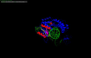
4QLB with conserved residues im pink
The VRR-Nuc containing proteins might present a single domain, only the VRR-Nuc domain [7], or present other domains, such as the human FAN1 (human FANCD2-associasted nuclease) [8] and its homologues [9]. The human FAN1 is composed by 4 well conserved domains: UBZ (ubiquitin-binding zinc finger), which participates in substrate recognition and binding; SAP (SAF-A/B, Acinus and PIAS) related to DNA binding; TRP (tetratricopeptide repeat) implicated in FAN1 dimerization and protein-protein interactions; and the VRR-Nuc catalytic domain. [10]
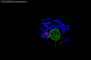
4R89 with conserved residues im pink and DNA strand in green
Alignment of the 4QLB structure with the 4R89. The conserved residues from the VRR-Nuc domains of both protein are marked in pink and present congruent positioning.
Enzymatic Activities
The VRR-Nuc domain can cleave the phosphodiesters bonds from nucleic acids. The domain in the single domain enzymes, such as psNUC from Psychrobacter (PDB:4qlb), presents Holliday Junction Resolvase activity [11]. The psNUC acts as dimer over four-way DNA-junctions, cleaving the DNA double-strand [12].
FAN1, however, is not able to process Holliday junctions, but has been identified presenting exo and endonuclease activity on different DNA substrates, such as: 5’ flaps; nicked double-strands; interstrand crosslinked (ICL) DNA; and double strands [13].
Biological functions and Related Pathogenesis
One of the first characterized VRR-Nuc containing proteins is the human FAN1. This enzyme is though to belong to a pathway of ICL DNA repair, which is fundamental for cell DNA integrity [14]. ICLs may occur from exposure of mutagenetic compound in the environment, such as chemotherapeutic treatments [15]. The ICLs are highly toxic since they hinder protein translation by preventing DNA strands separation [16]. FAN1 is capable of finding ICLs in the double stranded DNA and unhook the crossliked DNA, reaching the target by exo and endonuclease activity by the VRR-Nuc domain [17].
Fanconi Anemia (FA) is a genetic disorder caused by defects in one of the genes from the ICL repair pathway. The FAN1 was though to belong to this pathway for its direct interaction with the FANCD-2 protein, which coordinates proteins from the pathway. However, studies indicate that FAN1 acts independently of this pathway [18]. FAN1 defect leads to greater sensibility to ICL inducing agents, also confirming the enzyme in the ICL repair [19].


