Generalities
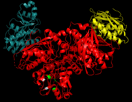
2x36 is a 6 chain structure with sequence from Human. This domain belongs to the Lon protease family.
Mitochondrial Lon protease is an ATP-dependent serine protease (an enzyme that hydrolyses proteins and polypeptides) involved in the selective degradation of misfolded proteins. LONP1 situated on chromosome 19 is the nuclear gene encoding mitochondrial Lon protein. The single species of mRNA of this protein is found in the mitochondrial matrix. This protein from human tissues has a molecular mass of 100 kDA.
It is involved in the selective degradation of misfolded proteins and also coordinates key processes through targeted proteolysis of folded proteins .Their only species of mRNA is found in the mitochondrial matrix and their proteolytic domain is the center of the Lon protease activity.
Function
The mitochondrial Lon protease is an important regulator of mitochondrial metabolism including the maintenance and repair of mitochondrial DNA thanks to its proteolytic domain. This protein is also essential for homeostasis of mitochondria, and by regulating some regulatory proteins which have a short life or damaged proteins. LONP1 works as a gatekeeper for specific proteins imported into the mitochondrial matrix. It also contributes to the degradation of imported, aberrant, unprocessed proteins using its protease activity.[1]
Lon protease has three main roles[2].
This protein is able to do a proteolytic digestion of oxidized proteins which allows the renewal of essential mitochondrial enzymes such as aconitase or Mitochondrial transcription factor A.
Lon protease is involved in mtDNA replication and mitogenesis by being a mitochondrial DNA-bing protein. Human Lon and mtDNA associate at the level of their at least 4 contiguous guanine sequence and form a G-quadruplex[3]. This G-rich region is the control region for mtDNA replication and transcription[4].
Mitochondrial Lon protease interacts with protein chaperone, notably HSP60-Hsp70 complex to protect cell from apoptosis under environmental stress[5].
The mitochondrial Lon protease is essentially found in the cytoplasmic of mitochondria because amino-acid has a potential mitochondrial targetting presequences[6].
Lon Human protease alternates between cycles of being bound to the mitochondrial genome and being free into the mitochondrial cytoplasm thanks to the proteolytic domain where it can degrade abnormal proteins coming from damaged proteins, errors in the synthesis, or misfolded of multimeric proteins. Its inactive conformation prevents uncontrolled proteolysis by the proteolytic domain.
To achieve proteolytic cleavage, the Lon protein has to form a hexamer.
Lon protease has also a role in mtDNA quality control by permits oxidative mitochondrial DNA damage. Sensitivities of H2O2-induced mtDNA damage depend on the proportion of LON[7].
Other ATP-dependent proteases are found in eukaryotic cells and organelles like 26S protease which uses ATP hydrolysis for conjugation or ubiquitin for example.
General structure
Lon proteins are grouped into two families, LonA and LonB. The human protein LonP1 is part of the LonA proteins [8]. This protein has three isoforms obtained by alternative splicing of the portion of DNA coding for this protein [9]: here is a possible cleavage at the amino and carboxyl ends ; thus, there is an isoform for each possible site of cleavage and one for both. [10]
Globally there is a great diversity of Lon proteins, but they are all organized in an oligomeric ring structure, mostly hexameric structure with identical subunits.
Lon proteins are therefore an hexameric chambered protease complex. (This structure is similar to yeast Pim1 )
The six Lon monomers are forming three pairs of legs owned by the N-terminal domain of the protein. This structure is emerging of the protein as a trimer of dimers [11].
Like many proteins, Lon is a flexible peptide which has different three-dimensional conformations. In the absence of substrate, Lon's hexameric ring adopts an open conformation, while upon substrate engagement, it changes into a closed but asymmetric ring conformation.
. The protein can therefore pass from one conformation to another by hydrolysis of ATP[12].
In the substrate-engaged form, sequential ATP hydrolysis drives the progressive translocation of unfolded substrates from the AAA+ channel towards the protease domains. With these conformational changes, the active sites of the Lon protein are protected from the external environment in the oligomeric complex that forms the degradation chamber. This mechanism is likely to be conserved in all Lon proteases and is related to the rotary treadmilling mechanism of other AAA+ translocases. The N-terminal domains show much larger sequence divergences across species and kingdoms than the A and P domains, and this is possibly related to organism-specific substrate recognition requirements. [13]
This form of degradation chamber is also found in bacteria, plants, fungi and metazoan, the similarities with bacteria are most probably due to the endosymbiotic theory.
This protein has a proteolytic and chaperone-like activity, it cannot unfold aggregated proteins, but can participate in the assembling of some complexes). These two enzymatic activities are separated on two polypeptide chains forming a complex or two separate domains on the same polypeptide chain.
The Lon protein has three main distinct domains: the first, the N-terminal domain, is specialised in substrate binding and oligomerization. The second, called the AAA+ domain (or A domain) corresponds to the fixation and hydrolysis site of the ATP, it also has unfolding activity. Finally, the third domain located at the C-terminal is an active serine site leading to substrate degradation. This is a proteolytic domain, called domain P [14].
Mammalian Lon protein only interacts with single-stranded DNA (ssDNA) but not dsDNA. There are therefore special sequences for interaction with G-rich DNA as well as RNA. In addition, the binding of a substrate to the protein stimulates the interaction with the DNA.
mtDNA binds to the Lon protein with different affinities depending on the state of the cell and the type of cell meeting the following four parameters [15]:
- the single stranding state of mtDNA
- the bioavailability of the mtDNA binding sites
- the affinity of the protein for a given DNA sequence
- the total number of high and low affinity Lon binding sites present
Structural highlights
From hLon main three domains, the ATPase domain and the C-terminal active site are those which confer to the protein its function.
ATP domain
The ATPase domain enables after the consumption of an ATP molecule to get the required energy for the active site to hydrolyze protein substrates. It has been demonstrated that the presence of ADP induces a conformational change to obtain an asymmetric hexametric ring. As a result, the catalytic site reaches its open state where the protein substrate can bind. ATP most likely replaces ADP from the ATPase domain to cut off the next substrate. In presence of AMP the hexametric ring takes a closed conformation state suggesting that until either ATP or ADP is present in the environment, hLon has the capacity to perform its catalytic activity[16].
Active site
The active site represent by 2x36 is composed of six protomers in the asymmetric unite. One protomer counts nine b-strands and seven a-helices. An analysis of the complex’ structure suggested that two pair of protomers form A:B and C:D dimers and that the two remaining ones remain uncoupled. The dimer interface A:B/C:D is mostly linked by one another through hydrophilic interactions, where the a1-helix is packed against the b3-strand and the loop between b7 and b8 makes inter-subunit contacts with b2[17].
As all LonA proteins, hLon catalytic activity relies on a Ser-Lys dyad. Ser855 on a2 conducts the catalytic cleavage with the assistance of Lys898 on a3 through their hydrogen-bonding. The lysine works as a general base along with Thr880 which, in their deprotonated form, abstract the proton from the nucleophilic serine. Those three residues constitute the . A characteristic of hLonP is that the 3(10) helix at the N-terminal end of a2 is able to bring an additional residue into the active site, Asp852. This most likely enables Lys898 pKa lowering by creating a hydrophobic environment, and thus, prevents the dyad to cut off protein substrates. This catalytic inactive form is also supported by the Asp852 and Trp770 residues that contribute to the . Asp852 removal from the active site through conformational changes enables hLon to reach an open state that can hydrolyze protein substrate through ATP consumption[18].
The figure below shows a 3D simulation of the 2x36 protein, in which the catalytic core (in green) and the catalytic site obstruction (in gray) are highlighted and can be more easily observed.

Evolutionary conservation
The Lon proteolytic domain has a highly conserved structure. Like its orthologues, namely the eubacterium E. coli (1rre), and the two archaea M. jannaschii and A. fulgidus, it presents at its C-terminal a Ser-Lys dyad responsible of the substrate degradation activity. Although hLonP active site resembles mostly to the one of EcLonP, the b5-sheet is replaced by an extension to a2. Thus, the N-terminal region of this helix carries the catalytic serine is a 3(10) helix and not a b-strand. As a consequence, hLonP has the ability to bring the Asp852 into the active site to close it by forming a hydrogen bond with Lys898, a property already observed in MjLon active site. This inactive state likely makes the catalytic serine inaccessible to the substrate and constraints the pKa of the lysine. Other main structural differences are loop shifts connecting the secondary structure elements b1 and b2, and a1 [19].
Disease
Various myopathy, type 2 diabetes, Parkinson's disease, or Alzheimer's disease are human degenerative disease partly due to abnormalities of the mitochondria[20]. In fact, Lon protease has a role in cancer, apoptosis and aging because this protein is an essential part of developmental pathways and stress response.
Mutant in Lon decreases the degradation capacity of proteins with abnormal conformations which lead to mitochondrial dysfunction. Mitochondrial dysfunction causes normal cells to become apoptotic, or to aberrant adaptation and selection of hypoxic phenotypes in pathological conditions like cancer[21].
Lon expression is necessary for survival in mammals. Indeed, a homozygous deletion of LONP1 is lethal for early embryonic[22]. Indeed, the CODAS Syndrome is a rare and multi-system developmental disorder from heterozygous or homozygous mutations in LONP1 where all the affected children were very severely impacted by their disease.
The LONP1 gene is regulated, when the cell undergoes a heat shock, starvation or oxidative stress the gene is up-regulated. On the contrary, Lon is down-regulated with aging, extensive hypoxia, and prolonged oxidative stress. So Lon is an important factor in aging and degenerative disease.
A consensus binding site of Nuclear Respiratory Factor 2 (NRF-2) is present on the region -623/+1 of the LONP1 promoter which is important for response to reactive oxygen species related to oxidative stress. As well as the putative binding site in -2023/-1230 for NF-kB in LONP1 which consolidate the role of Lon as a stress protein[23].
Research is being done to use Lon as a therapeutic target for the treatment of cancer by developing novel Lon inhibitors.

