Introduction
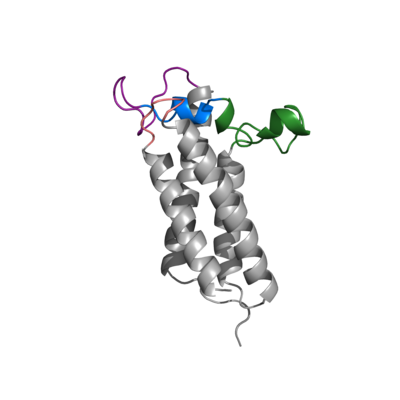
Figure 1. Closed Conformation of VKOR due to Warfarin Binding
Vitamin K Epoxide Reductase (VKOR) is an endoplasmic membrane enzyme that generates the active form of Vitamin K to support blood coagulation. The vitamin K Cycle, and the VKOR enzyme specifically are common drug targets for thromboembolic diseases. This is because, as pictured, the vitamin K cycle
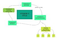
Figure 2. Overview of Vitamin K Cycle
is the process in which blood coagulant factors II, VII, IX, and X are activated. This promotes blood clotting, which can be dangerous and cause thromboembolic diseases such as stroke, deep vein thrombosis, and/or pulmonary embolism.
Location of Enzyme
Vitamin K Epoxide Reductase is found and primarily synthesized in the liver. It is embedded in the membrane known as the endoplasmic reticulum.
Structure
VKOR WIKI
The VKOR enzyme is made up of four transmembrane helices: T1, T2, T3, and T4.(Pictured in Grey) Each of these helices come together to form a hydrophobic pocket, that is topped by a cap domain. In the cap domain are important regions that are significant for Vitamin K binding, and the overall function of Vitamin K Epoxide Reductase. These important regions are the Anchor(Green), Cap Region (Blue), Beta Hairpin (Purple), and 3-4 Loop (Pink).
[1]
Transmembrane Helices
Cap Domain
Significant Cysteines
Vitamin K Epoxide
Warfarin


