Background
The anaplastic lymphoma kinase (ALK) was first discovered in 1994 as a tyrosine kinase in anaplastic large-cell lymphoma (ALCL) cells.[1] The specific type of tyrosine kinase ALK is classified as is a receptor tyrosine kinase (RTK) and like other RTKs, it's an integral protein with extracellular and intracellular domains and is involved in transmembrane signaling and communication within the cell. ALK is commonly expressed in the development of the nervous system. Anaplastic lymphoma kinase receptor (ALKr) is the extracellular portion of the RTK that includes a binding surface for a ligand to bind. When the ALK activating ligand (ALKAL) binds to ALKr, this causes a conformational change of ALK, allowing two ALK-ALKAL complexes to interact with each other, which will then allow intracellular kinase domain of ALK to phosphorylate a tyrosine residue on a downstream enzyme, which will activate this enzyme and activate a signaling cascade. Abnormal forms of ALK are closely related to the formation of several cancers. [2]
Structure & Function
Domains
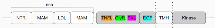
Figure 1: Anaplastic Lymphoma Kinase and its domains.
The region from NTR to the MAM is the Heparin Binding Domain. The TNFL-PXL are the extracellular domains and the EGF is the domain that binds the extracellular region with the extracellular region of the transmembrane. The TMH is the transmembrane domain. The kinase domain is the intracellular portion of the ALK. The extracellular portion of ALK has an inactive state, which is its monomerized form, and an active dimerized state with its ligands bound. The monomer is shown in Figure 2, which has many different domains (Figure 1). The growth factor-like domain (EGF) connects the extracellular domains to the transmembrane domain (cyan). The tumor necrosis factor-like domain (TNFL) has a beta-sandwich structure that provides important residues that act as the binding surface for the ligand (orange). The glycine-rich domain (GlyR) contains 14 rare polyglycine helices that are hydrogen-bound to each other (green). The of these rare helices create a very rigid structure that is important for ALK function. The polyglycine extension loop (PXL) connects two of these polyglycine helices (pink).
The domains that aren't shown in Figure 2 but are shown in the domain map (Figure 1) also make up the monomer. The heparin binding domains (HBDs), are at the N-terminal end of the monomer. Heparin has been found to be a possible activating ligand of ALK.[3] The transmembrane domain (TMH) contains the residues of ALK that are located within the membrane. The kinase domain is the intracellular portion of ALK that contains the Tyr residues which are auto-phosphorylated when ALK is activated, initiating a signaling cascade. [4]
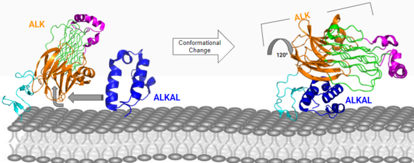
Figure 3: ALK-ALKAL complex, showing the conformation change of ALK from the binding of ALKAL.
PDB: 7N00 Conformational Change
The anaplastic lymphoma kinase activating ligand (ALKAL) binds to the on the ALK at the TNFL domain. This induces a conformational change which allows for the PXL and the GlyR domains to hinge forward.[5] (Figure 3) ALK's TNFL has E978, E974, E859, and Y966 that form salt bridges with R123, R133, R136, R140, and R117 on ALKAL that allow for activation, leading to of two ALK-ALKAL monomers.
Membrane Guidance of ALKAL to ALK
The negatively charged phosphate groups on the cell membrane interact with a highly conserved positively charged on ALKAL that faces the membrane. These (7MZZ) guides ALKAL to ALK and correctly positions ALKAL for its binding surface to face ALK's binding surface, which allows for a more favorable interaction.
Role of Activated ALK
Once the ALKAL binds with ALK and dimerizes with another ALK-ALKAL complex, this activated conformation also initiates a conformational change of the intracellular kinase domain of ALK. This causes an autophosphorylation of several tyrosine residues of this domain, activating a signaling cascade with its kinase activity.
Disease
There are many that would cause constitutive receptor activation, enhancement between the interaction of receptors or stabilization of active receptors are known to relate to oncogenic potentials (Figure 4). The that is mutated to an arginine is known to be a gain-of-function in lung adenocarcinoma which can lead to constitutive activation of ALK. The that is mutated to the a serine may cause a gain-of-function mutation that is linked to acute myeloid leukemia. When the is mutated to a glutamate it is commonly identified in histiocytic neoplasms. The changing to arginine could cause possible oncogenic potentials which are not specified yet. The F856S and R753Q mutations are known to increase cytokine-dependent cell proliferation in certain cells. [6]
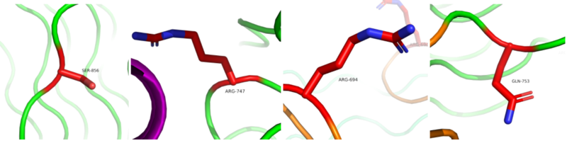
Figure 4: Mutated residues on ALK that contribute to stabilization of the active state of ALK, leading to many types of cancers. From left to right: F856S, G747R, H694R, R753Q
PDB: 7N00


