This old version of Proteopedia is provided for student assignments while the new version is undergoing repairs. Content and edits done in this old version of Proteopedia after March 1, 2026 will eventually be lost when it is retired in about June of 2026.
Apply for new accounts at the new Proteopedia. Your logins will work in both the old and new versions.
1gux
From Proteopedia
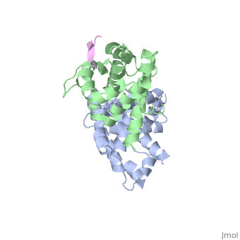

RB POCKET BOUND TO E7 LXCXE MOTIF
Overview
The pocket domain of the retinoblastoma (Rb) tumour suppressor is central to Rb function, and is frequently inactivated by the binding of the human papilloma virus E7 oncoprotein in cervical cancer. The crystal structure of the Rb pocket bound to a nine-residue E7 peptide containing the LxCxE motif, shared by other Rb-binding viral and cellular proteins, shows that the LxCxE peptide binds a highly conserved groove on the B-box portion of the pocket; the A-box portion appears to be required for the stable folding of the B box. Also highly conserved is the extensive A-B interface, suggesting that it may be an additional protein-binding site. The A and B boxes each contain the cyclin-fold structural motif, with the LxCxE-binding site on the B-box cyclin fold being similar to a Cdk2-binding site of cyclin A and to a TBP-binding site of TFIIB.
About this Structure
1GUX is a Protein complex structure of sequences from Homo sapiens and Human papillomavirus. Full crystallographic information is available from OCA.
Reference
Structure of the retinoblastoma tumour-suppressor pocket domain bound to a peptide from HPV E7., Lee JO, Russo AA, Pavletich NP, Nature. 1998 Feb 26;391(6670):859-65. PMID:9495340 Page seeded by OCA on Fri May 2 18:02:15 2008
