|
Structure
Syncytin is a human endogenous envelope protein and is found in the human placenta. There are two forms of syncytin; syncytin 1 and syncytin 2, and they are both critical for fetal implantation and placental development. Syncytin 1 contains 538 amino acids and is located on the human chromosome 7 while the specific HERV-W1 coding region is located at 7q21.2. Syncytin has two domains, the surface unit and the transmembrane unit. The surface unit of syncytin 1 is what binds to receptors on the host cell whereas the transmembrane unit supports the fusion of two cells [1]. The structure of syncytin is composed of three identical monomers and the precursor that synthesizes the formation is glycosylated gPr73. Once the precursor is cleaved at the surface unit and the transmembrane unit, both units covalently bond via disulfide binding. The transmembrane unit is significant as it contains immunosuppressive domain known as the fusion peptide that plays a critical role in the tolerance a mother has to her fetus during pregnancy [2]. Thus, the mother’s immune system does not “attack” the fetus as if it is a foreign body.
Function
Syncytin-1 and Syncytin-2 are both fusogenic which means they facilitate the fusion of cells. Together, the proteins are required for the proper placental formation and maintenance [2]. The of the membrane occurs after conformational changes occur within the surface unit and the transmembrane unit. For these changes to occur, the surface unit has to bind with specific cells to release the “cap” which then opens up the space of the transmembrane unit to be exposed. This then creates a hairpin that then allows for the fusion of the two membranes together as it brings them close enough for the interaction to occur. In order for this to occur, the fusion of cells together creates syncytiotrophoblasts and these structures allow for the exchange of maternal blood with fetal blood and ultimately establish the flow of nutrients, oxygen transport, and hormone synthesis as well [3]. This allows for the fetus to grow and develop. However, when unregulated or in excess, Syncytin-1 has been linked to several diseases. The increased inflammatory response has been linked to individuals with a higher level of Syncytin-1 which has been shown to increase the likeliness of developing neuropsychological disorders [1].
Evolutionary Relevance
8% of the human genome consists of human endogenous retroviruses and these human endogenous retroviruses have been linked to the early evolution of primates due to germline infections of exogenous retroviruses. These retroviruses consist of the genes gag, pro, pol, and env. The env gene is associated with the envelope of the retrovirus and has been linked to prompting cancerogenesis and the gag and pol genes have been linked to the inactivation of mutations. Syncytin-1 has been found to have been conserved throughout hominoids and has maintained fusogenic activity. The gene itself has become a key factor for reproduction in mammals. This implies that Syncytin-1 has been co-opted by hominoids for placental development as well and the proper blood barrier needed to supply the fetus with nutrients and other materials needed for the growth and development of the fetus. The immunosuppression of the mother’s immune system plays a critical role in the survival of the fetus and its development. This increases the viability of pregnancy and decreases the chance of pregnancy loss which is beneficial to mammalian species [4].
Disease
Multiple Sclerosis (MS)
An increase in the expression of Syncytin-1 has been directly correlated to the development of multiple sclerosis. It was also found that syncytin-1 could induce inflammation and was found to cause neurotoxic cascades as well. This neurotoxic cascade includes the secretion of proinflammatory cytokines. Multiple sclerosis is an inflammatory disorder that causes the demyelination of axons. It was found that overexpression of Syncytin-1 can cause tissue damage and demyelination if affecting glial cells. Overexpression of Syncytin-1 in immune cells that are activated in multiple sclerosis and patients that are affected with acute infections have an elevated level of monocytes that contain the protein. Along with this, after interaction with lipopolysaccharides, the level of syncytin-1 also increased in T and B lymphocytes along with natural killer cells which implies that syncytin-1 may have a role in immune cell activation in the early stages and may even be involved in the pathogenesis of multiple sclerosis [1].
Schizophrenia
The overexpression of Syncytin-1 has also been loosely correlated to disorders such as schizophrenia. Brain-derived neurotrophic factors, neurotrophic tyrosine kinase receptor types, and dopamine receptor D3 are all significant in the development of schizophrenia, and it was found that the overexpression of the protein in human U251 glioma cells correlates to the increase of these receptors [2]. Studies have shown that an increase in the transcription of syncytin-1 is also correlated to the development of schizophrenia. However, several studies have also shown that there is no significant difference in the expression level of syncytin-1 in the development of schizophrenia [1]. However, these findings imply that syncytin may have a variety of different ways of affecting the development of schizophrenia.
Alzheimer's Disease
An abnormal influx of calcium has been linked to the development of Alzheimer’s Disease in individuals. Overexpression of syncytin-1 has been linked to the influx of calcium ions in neuroblastoma cells. Along with the influx of calcium, nitric oxide also contributes to the development of Alzheimer’s Disease as it regulates the inflammatory response of the brain and neuronal cells [2]. An increase in syncytin-1 levels has the capability to activate the enzyme nitric oxide synthase |
Nitric Oxide Synthase (NOS) or Nitric Oxide Synthase oxygenase is an enzyme catalysing the formation of L-citrulline and nitric oxide (NO) from L-arginine. NOS is a homodimeric protein with 125- to 160-kDa per monomer. In mammals, NOS appears as 3 isozymes: neuronal NOS (nNOS) (for details see Nos1), cytokine-inducible NOS (iNOS) and endothelial NOS (eNOS). The N-terminal domain of NOS is an oxygenase domain (OD). NOS cofactors are: NADPH, FAD, FMN, heme and O2. See also Nos1.
Introduction
In mammals three isozymes of NOS have been identified: Neuronal NOS (nNOS), inducible NOS (iNOS), and endothelial NOS (eNOS)[6]. (The NOS enzymes are found in numeral organisms. Most facts used here are from the human NOS, but sites from different organisms are used). Neuronal NOS is producing NO in the nervous tissue in both the peripheral and the central nervous system. nNOS is functioning in cell signaling and communication - a vital part of the nervous tissue. Inducible NOS is connected with the immune system. Endothelial NOS is controlling the amount of NO signaling in the endothelial cells eg. blood vessel dilation. An overview of the structural organization of the NOS homodimer is given below. All cofactors are included and the electron transfer pathway which takes place in NOS is indicated.
Each NOS monomer is composed of two domains: an N-terminal oxygenase domain and a C-terminal reductase domain. Each subunit is held together by a zinc ion, which is bound by two cysteines from each oxygenase domain. Calmodulin binds to a linker region between the two domains and is necessary for activity. The reductase domain supplies electrons for the NOS reaction which takes place in the oxygenase domain. The reductase domain contains two redox-active prosthetic groups, flavin adenine dinucleotide (FAD) and Flavin mononucleotide (FMN). Nicotinamide adenine dinucleotide phosphate(NADPH) binds to the domain and passes on an electron to FAD which passes the electron on to FMN. FMN passes the electron on to the heme in the oxygenase domain on the opposite subunit. The oxygenase domain contains 5,6,7,8-tetrahydrobiopterin (H4B) and the already mentioned heme. These two are also redox active groups. Besides Heme and H4B, the oxygenase domain binds the substrate L-arginine which takes part in the NO synthase reaction (see below).
Many structures of NOS have been determined. A list with additional information can be seen here.
The reaction of NOS
As previously described NOS is an enzyme consisting of two domains; the N-terminal oxygenase domain and the C-terminal reductase domain.
The NO production takes plase in the oxygenase domain, whereas the reductase domain provides the electrons necessary to drive the reaction in the oxygenase domain.
The reaction is:
The reaction occurs in two steps. In the first step L-arginine is turned in to N-hydroxy-arginine. In the second step L-citrulline and NO are formed. The reaction utilizes 2O2 and 3 electrons from 3/2 NADPH.[7]
The Oxygenase domain of NOS
The contains the active site of the enzyme. The active site binds the substrate (Arginine) which is converted into citruline and NO (explained in details below). The domain has three cofactors bound:
((6R).5,6,7,8-Tetrahydrobiopterin)
(Heme)
General Structure
The oxygenase domain can be divided into three subdomains. Firstly, the
, which is crescent in shape, binds the substrate in an interior pocket of the crescent. The Heme group is located between the tips of the crescent shape, thus closing the pocket in which the substrate is located. Secondly the serves as a cap for the cavity created by the substrate binding domains crescent shape. Thirdly there is a subdomain consisting of making up a hydrophobic core, however this subdomain is not thought to take part in the catalytic activity of the enzyme.
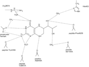
Substrate binding
The active site is highly conserved in the different NOS species. Thus it is possible to discuss substrate binding i general terms. The NOS enzyme binds its substrate (L-arginine) in the distal pocket by hydrogen bindings both to the guanidino[1] end and the amino acid end. is shown in green with the heme and H4B shown. NOS binds its substrate by coordinating CO(or O2) to the heme at the site occupied by oxygen[8](it is the opposite site of the Cys coordination to heme - look in the 'heme' section). The binding of substrate leads to a 2-step transformation first to N-hydroxy-L-arginine (the tightly bound intermediate) and then NO and L-Citrulline. The product NO can then either diffuse out of the or bind to the heme and function in NO auto-inhibition though this inhibition is diverse throughout the 3 isoforms[9].
PDB structures used in the section above: 3nos, 2g6h
H4B
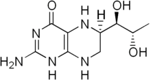 Structure of tetrahydrobiopterin is an essential cofactor in NOS and in the aromatic amino acid hydroxylases. NOS contains two molecules of , one in each monomer. The active site forms part of the cavity already described. This cavity can be visualized as a . Here H4B helps substrate interactions by lining the active-center tunnel and hydrogen bonding to the heme propionate and to alfa helix 7. The propionate group and alpha helix 7 are also involved in the L-Arg binding. This gives H4B the opportunity to play an important role in the control of subunit interactions and active site formation. H4B is therefore more or less a structural cofactor and has a stabilizing effect. Its structural importance is further reconned to play a role in dimer formation (dimerization requires bound zinc ion along with H4B), and major conformational changes leading to the formation of the active site channelform[10].
 Hydrogenbondings in H 4B binding site ( 2nse) The H4B is bound by H-bonds to several of the residues surrounding it. For example is O4 of H4B H-bonded to Arg 367 and H4B N3 is H-bonded to one of the heme propionate groups ( the heme propionate group has two carboxylate oxygens in use for H-bonds)[10]. The overall picture of all the H-bonds can be seen by clicking on the figure on the left.
But H4B is not only a structural cofactor, it also plays a very important role in NO synthesis, donating an electron to the heme.[11] H4B can deliver an electron to the heme much faster than the reductase domain can, therefor H4B is used by NOS in the Arg hydroxylation, activating O2 by providing the second electron. Thus, H4B is a kinetically prefered electron donor. As shown in the reaction (bottom right, click for enlargement) the second electron, that H4B donates, helps the FeIIO2 intermediate to be reduced to oxidants that are able to react with Arg and N-hydroxy-L-arginine (NOHA) [11] If H4B was not present the FeIIO2 intermediate would decay to superoxide and ferric enzyme due to the reductase domain being slower to deliver an electron than the proces of decay is to happen. H4B is faster than both of these processes[11].
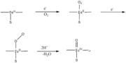 model for NOS oxygen activation PDB structures used in the section above: 3nos, 2g6h
Heme
Heme is a prosthetic group containing an iron ion in the center of a large organic ring called porphyrin (See picture). The heme group can have several functions: it can act as a transporter of diatomic gases, as a chemical catalyst, detect diatomic gases, and take part in electron transfer. In NOS the heme both binds a diatomic molecule (CO, NO, O2) and functions in the multi electron pathway [12].
As mentioned, the heme group takes part in the creation of the cavity running through the monomer. Arginine/citrulline diffuses in and out this cavity. The heme group in the cavity is held in place by van der Waals interactions with hydrophobic and aliphatic side chains of the protein making up the cavity. The propionic acid groups of heme forms several hydrogen bonds with water molecules inside the cavity. The iron in heme in pentacoordinated, with its axial ligand supplied by S of cysteine 186 (2nse) [13].
Zinc
In order for NOS to be active it has to dimerize and bind H4B. The dimer is structurally stabilized by a .
which is situated at the oxygenase domain interface of the dimer[13]. The zinc ion is tetrahedrally coordinated by four cysteins (two from each monomer - Cys109 and Cys104). The zinc ion is found at a region which connects the N-terminal hook and the subunit core. The coordination of zinc arranges the N-terminal hooks so that they interact with their own subunit. However, when there is no zinc ion present, two of the thiolate ligands (cysteines) form a disulfide bond connecting the two subunits[14].
PDB structures used in the section above: 2g6h
The Reductase Domain of NOS
The reductase domain of the NOS homodimer will not be discussed thoroughly on this page. However, a short discussion of the electron transfer which occurs will be given along with an introduction to the bound cofactors and the general structure.
The has three cofactors bound.
(Nicotinamide adenine dinucleotide phosphate)
(Flavin adenine dinucleotide)
(Flavin mononucleotide)
The reductase domain is, as mentioned, bound to the oxygenase domain by a linker region that binds calmodulin. The calmodulin linker consists of 32 residues. The binding of Ca2+ loaded calmodulin to the linker region is found to be crucial in that it induces a conformational change which is essential for electron transfer. The NOS activity is thereby dependent upon the Ca2+ concentration It is important to emphasize that the electron transfer occurs from the reductase domain of one subunit to the oxygenase domain of the opposite subunit (i.e. a trans transfer). The conformational change induced by calmodulin binding brings the mentioned reductase and oxygenase domains closer together, thus the linker acts as a hinge. The electron transfer occurs two times per NO molecule produced. The first transfer supplies an electron for the conversion of L-arginine to its intermediate, the second transfer for the conversion of the intermediate Citrulline and NO. In general the reductase domain can be divided into three subdomains: the NADPH binding domain, the FAD binding domain, and the FMN binding domain. The NADPH and FAD binding domains are associated whereas the FAD and FMN domains are connected by an α-helical binding domain. The electrons donated by NADPH is passed on to FAD. FAD then shuttles the electron to FMN. The FMN binding domain is a flexible domain and here the conformational change occurs. The binding of calmodulin rotates the reductase domain and oxygenase domain along a vertical axis, thus bringing the reductase domain closer to the opposite oxygenase domain. The electron can then due to shorter distance be passed on the the heme group of the oxygenase domain [15]. The iron ion in the heme group is reduces from iron (III) to iron (II) which catalyses the substrate reaction.
Structures used in the section above: 1tll
3D Structures of Nitric Oxide Synthase
Nitric Oxide Synthase 3D structures
|
References
- ↑ 1.0 1.1 1.2 1.3 1.4 Wang, X., Huang, J., & Zhu, F. (2018, September 7). Human endogenous retroviral envelope protein syncytin-1 and inflammatory abnormalities in neuropsychological diseases. Frontiers in psychiatry. Retrieved April 18, 2022, from https://www.ncbi.nlm.nih.gov/pmc/articles/PMC6137383/
- ↑ 2.0 2.1 2.2 2.3 2.4 Durnaoglu, S., Lee, S.-K., & Ahnn, J. (2021). Syncytin, envelope protein of human endogenous retrovirus (Herv): No longer ‘fossil’ in human genome. Animal Cells and Systems, 25(6), 358–368. https://doi.org/10.1080/19768354.2021.2019109
- ↑ Ruigrok, K., Vaney, M.-C., Buchrieser, J., Baquero, E., Hellert, J., Baron, B., England, P., Schwartz, O., Rey, F. A., & Backovic, M. (2019, November 8). X-ray structures of the post-fusion 6-helix bundle of the human Syncytins and their functional implications. Journal of Molecular Biology. Retrieved April 18, 2022, from https://www.sciencedirect.com/science/article/pii/S0022283619306163
- ↑ Cáceres, M., & Thomas, J. W. (2006). The gene of retroviral origin syncytin 1 is specific to hominoids and is inactive in Old World Monkeys. Journal of Heredity, 97(2), 100–106. https://doi.org/10.1093/jhered/esj011
- ↑ Locke, W. J., Guanzon, D., Ma, C., Liew, Y. J., Duesing, K. R., Fung, K. Y. C., & Ross, J. P. (2019, November 14). DNA methylation cancer biomarkers: Translation to the clinic. Frontiers. Retrieved April 27, 2022, from https://www.frontiersin.org/articles/10.3389/fgene.2019.01150/full#:~:text=Hypermethylation%20can%20drive%20the%20silencing,et%20al.%2C%202015).
- ↑ Flemming, I., Chapter 3, Biology of Nitric Oxide Synthases, from Microcirculation, Editors Ronald F. Tuma, R. F., Durán, W. N., and Ley, K., 2nd edition, Academic Press, 2008
- ↑ Gorren AC, Mayer B. Nitric-oxide synthase: a cytochrome P450 family foster child. Biochim Biophys Acta. 2007 Mar;1770(3):432-45. Epub 2006 Sep 1. PMID:17014963 doi:10.1016/j.bbagen.2006.08.019
- ↑ Fan B, Wang J, Stuehr DJ, Rousseau DL. NO synthase isozymes have distinct substrate binding sites. Biochemistry. 1997 Oct 21;36(42):12660-5. PMID:9376373 doi:http://dx.doi.org/10.1021/bi9715369
- ↑ Rousseau DL, Li D, Couture M, Yeh SR. Ligand-protein interactions in nitric oxide synthase. J Inorg Biochem. 2005 Jan;99(1):306-23. PMID:15598509 doi:http://dx.doi.org/10.1016/j.jinorgbio.2004.11.007
- ↑ 10.0 10.1 Raman CS, Li H, Martasek P, Kral V, Masters BS, Poulos TL. Crystal structure of constitutive endothelial nitric oxide synthase: a paradigm for pterin function involving a novel metal center. Cell. 1998 Dec 23;95(7):939-50. PMID:9875848
- ↑ 11.0 11.1 11.2 Wei CC, Wang ZQ, Meade AL, McDonald JF, Stuehr DJ. Why do nitric oxide synthases use tetrahydrobiopterin? J Inorg Biochem. 2002 Sep 20;91(4):618-24. PMID:12237227
- ↑ Li H, Igarashi J, Jamal J, Yang W, Poulos TL. Structural studies of constitutive nitric oxide synthases with diatomic ligands bound. J Biol Inorg Chem. 2006 Sep;11(6):753-68. Epub 2006 Jun 28. PMID:16804678 doi:http://dx.doi.org/10.1007/s00775-006-0123-8
- ↑ 13.0 13.1 Fischmann TO, Hruza A, Niu XD, Fossetta JD, Lunn CA, Dolphin E, Prongay AJ, Reichert P, Lundell DJ, Narula SK, Weber PC. Structural characterization of nitric oxide synthase isoforms reveals striking active-site conservation. Nat Struct Biol. 1999 Mar;6(3):233-42. PMID:10074942 doi:http://dx.doi.org/10.1038/6675
- ↑ Crane BR, Rosenfeld RJ, Arvai AS, Ghosh DK, Ghosh S, Tainer JA, Stuehr DJ, Getzoff ED. N-terminal domain swapping and metal ion binding in nitric oxide synthase dimerization. EMBO J. 1999 Nov 15;18(22):6271-81. PMID:10562539 doi:http://dx.doi.org/10.1093/emboj/18.22.6271
- ↑ Garcin ED, Bruns CM, Lloyd SJ, Hosfield DJ, Tiso M, Gachhui R, Stuehr DJ, Tainer JA, Getzoff ED. Structural basis for isozyme-specific regulation of electron transfer in nitric-oxide synthase. J Biol Chem. 2004 Sep 3;279(36):37918-27. Epub 2004 Jun 17. PMID:15208315 doi:http://dx.doi.org/10.1074/jbc.M406204200
in astrocytes which then induces the release of the redox reaction product, nitric oxide. These, in turn, are cytotoxic to oligodendrocytes which can damage axons [1].
Cancer
Syncytin-1 has been correlated to several different kinds of cancers including breast cancer, carcinomas, and endometrial cancers. As mentioned above, the env gene of syncytin-1 can become activated and promote the production of cancerous cells [2]. Overexpression in syncytin-1 contributes to increased proliferation, metastasis, and the growth of tumors in individuals due to the methylation of DNA and the 5’LTR of the syncytin gene. The overmethylation contributes to the silencing of the tumor-suppressing gene [5]. As a result, if both tumor suppressor genes in a cell contain a mutation, then the suppressor gene is deactivated. With the tumor suppressor gene losing function, cells may grow and divide unregulated, resulting in cancer.
|