We apologize for Proteopedia being slow to respond. For the past two years, a new implementation of Proteopedia has been being built. Soon, it will replace this 18-year old system. All existing content will be moved to the new system at a date that will be announced here.
ATPase
From Proteopedia
| |||||||||||
Contents |
3D Printed Physical Model of ATP Synthase
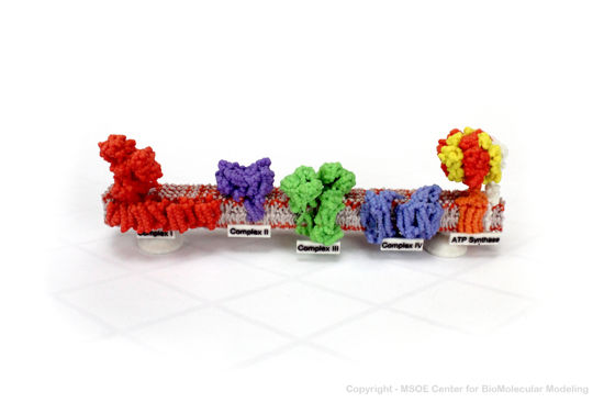
Shown below is a 3D printed physical model of the Respiration Electron Transport Chain. Complex I is colored red, complex II is purple, complex III is green, complex IV is blue and the atp synthase protein is colored orange, yellow and red.

The MSOE Center for BioMolecular Modeling
The MSOE Center for BioMolecular Modeling uses 3D printing technology to create physical models of protein and molecular structures, making the invisible molecular world more tangible and comprehensible. To view more protein structure models, visit our Model Gallery.
3D Structures of ATPase
References
- ↑ Rappas M, Niwa H, Zhang X. Mechanisms of ATPases--a multi-disciplinary approach. Curr Protein Pept Sci. 2004 Apr;5(2):89-105. doi: 10.2174/1389203043486874. PMID:15078220 doi:http://dx.doi.org/10.2174/1389203043486874
- ↑ Neupane P, Bhuju S, Thapa N, Bhattarai HK. ATP Synthase: Structure, Function and Inhibition. Biomol Concepts. 2019 Mar 7;10(1):1-10. doi: 10.1515/bmc-2019-0001. PMID:30888962 doi:http://dx.doi.org/10.1515/bmc-2019-0001
Proteopedia Page Contributors and Editors (what is this?)
Michal Harel, Wayne Decatur, Alexander Berchansky, Mark Hoelzer, Marius Mihasan, Karsten Theis, Jaime Prilusky

