We apologize for Proteopedia being slow to respond. For the past two years, a new implementation of Proteopedia has been being built. Soon, it will replace this 18-year old system. All existing content will be moved to the new system at a date that will be announced here.
Sandbox Reserved 1789
From Proteopedia
This page, as it appeared on June 14, 2016, was featured in this article in the journal Biochemistry and Molecular Biology Education.
Contents |
SHOC2-PP1C-MRAS
Introduction
Receptor Tyrosine Kinase Receptor
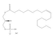
Lysophosphatidic Acid
Overall Structure
| |||||||||||
Student Contributors
Madeline Gilbert Inaya Patel Rushda Hussein


