Structure
There are three main domains of the Thyroid Stimulating Hormone Receptor. First is the shown in green. Region is concave in shape and is made up of primarily beta sheets and rich in Leucine. It is also called the leucine rich region. This is also the domain for key Lysine residues, which play a key role in binding. Second is the shown in pink. This domain is made up of seven helices and undergoes a conformation change upon ligand binding that activates the GPCR signal cascade (GPCR is shown in yellow). The Third region of the TSHR is the shown in orange. The Hinge Region plays a key role in the movement and stability of the TSHR.
Binding of Thyroid Stimulating Hormone to TSHR
The Thyroid Stimulating Hormone by complementary shape. The ECD and is curved and compliments the curvature of TSH similar to how a baseball fits into a glove. There are also several key ionic interactions between the TSH and TSHR. The key ionic interactions occurs in the which is highlighted in yellow. The seatbelt region is located in the beta subunit of the TSH. is Glu118 from TSH and Lys58 from the ECD. is between Asp111 from the TSH and Lys209 from the ECD. These interactions form salt bridges between the ECD and the TSH which allows for specificity of binding for TSH to TSHR.
Other key interactions that allow for specificity of binding are between the helix 1. Helix 1 contains several polar residues that interact with surrounding nonpolar residues like Leu62, Arg54, and Phe17. These interactions increase the activation potency and helps activate the push and pull mechanism of the hinge region.
Blocking TSHR in Active/Inactive States
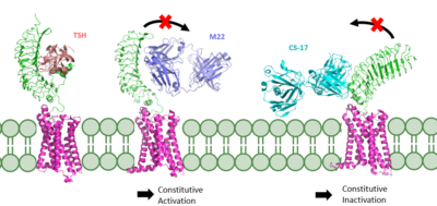
TSHR in active and inactive binding states.
Left is TSH bound to TSHR.
Middle is M22 bound to TSHR.
Right is CS-17 bound to TSHR.
The interactions between the ligand and the receptor have important consequences for disease states. In the image shown to the right are three different states of TSHR. The right-most structure is TSH bound to TSHR and is in it's upright active conformation. Comparing this is M22, shown in the middle, TSH binding offer not steric hinderance to keep it from being released. M22 can manifest with elevated levels of thyroid hormones which could be found in a person with Graves disease. M22 bound to TSHR is in the upright state and prevents transition to the down state because of the steric clash with the membrane which causes constitutive activation and symptoms of Graves disease. On the right most side is CS-17 bound to the TSHR. In contrast to TSH and M22 binding, CS-17 binds and locks TSHR in the down, inactive conformation. This prevents the signaling cascade to translation and causes constitutive inactivation.
These different ways to active and inactive TSHR could represent potential therapies for someone with graves disease or other thyroid related diseases with overacting TSH binding. Whereas current therapies target T3/T4 synthesis or destroy the gland using artificial hormones. These diseases could instead be targeted with something like M22 or CS-17
Structural highlights

