Structural highlights
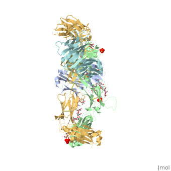
5dk3 is a 4 chain structure with sequence from Homo sapiens. Full crystallographic information is available from OCA. For a guided tour on the structure components use FirstGlance.
|
| Method: | X-ray diffraction, Resolution 2.28Å |
| Ligands: | , , , , , , , |
| Resources: | FirstGlance, OCA, PDBe, RCSB, PDBsum, ProSAT |
Publication Abstract from PubMed
Immunoglobulin G4 antibodies exhibit unusual properties with important biological consequences. We report the structure of the human full-length IgG4 S228P anti-PD1 antibody pembrolizumab, solved to 2.3-A resolution. Pembrolizumab is a compact molecule, consistent with the presence of a short hinge region. The Fc domain is glycosylated at the CH2 domain on both chains, but one CH2 domain is rotated 120 degrees with respect to the conformation observed in all reported structures to date, and its glycan chain faces the solvent. We speculate that this new conformation is driven by the shorter hinge. The structure suggests a role for the S228P mutation in preventing the IgG4 arm exchange. In addition, this unusual Fc conformation suggests possible structural diversity between IgG subclasses and shows that use of isolated antibody fragments could mask potentially important interactions, owing to molecular flexibility.
Structure of full-length human anti-PD1 therapeutic IgG4 antibody pembrolizumab.,Scapin G, Yang X, Prosise WW, McCoy M, Reichert P, Johnston JM, Kashi RS, Strickland C Nat Struct Mol Biol. 2015 Dec;22(12):953-8. doi: 10.1038/nsmb.3129. Epub 2015 Nov, 23. PMID:26595420[1]
From MEDLINE®/PubMed®, a database of the U.S. National Library of Medicine.
See Also
References
- ↑ Scapin G, Yang X, Prosise WW, McCoy M, Reichert P, Johnston JM, Kashi RS, Strickland C. Structure of full-length human anti-PD1 therapeutic IgG4 antibody pembrolizumab. Nat Struct Mol Biol. 2015 Dec;22(12):953-8. doi: 10.1038/nsmb.3129. Epub 2015 Nov, 23. PMID:26595420 doi:http://dx.doi.org/10.1038/nsmb.3129