This is a default text for your page '. Click above on edit this page' to modify. Be careful with the < and > signs.
You may include any references to papers as in: the use of JSmol in Proteopedia [1] or to the article describing Jmol [2] to the rescue.
Introduction
SARS-COV-2 Spike Protein
The of SARS-COV-2 is a symmetric trimer featuring 3 spike glycoprotein chains (UNIPROT: P0DTC2). Each monomer of the spike is called a spike glycoprotein, and the total assembly contains 2 main parts: The and subunits[3]. However, the native spike protein does not exist in this state prior to infection. The protein is actually inactive initially, but is later activated by proteases cleaving the inactive S protein into its two active subunits[3]. The S1 subunit contains the . The RBD is responsible for binding to the ACE2 receptor on the surface of the target cell, as well as neutralizing antibodies. The NTD, CTD, and their relevant interfaces actually play much larger roles in the binding of the spike protein to ACE2 than the RBD does due to their larger surface areas[3]. The S2 subunit is responsible for viral fusion and entry. Once bound to ACE2, and after the different domains in S2 have anchored to the membrane as well as delivered the viral envelope, the S2 subunit then changes conformation from the pre-hairpin to postfusion-hairpin conformation[3]. The S2 subunit contains a fusion peptide domain (FP), heptapeptide repeat sequences 1 and 2 (HR1 & HR2), TM domain, and cytoplasmic fusion domain (CT). Full information about the location and structures of these domains within the S2 subunit can be found in references 1 and 3[3][4]. For the purpose of this article about the minibinders, attention will be directed to the S1 subunit and its binding properties with ACE2.
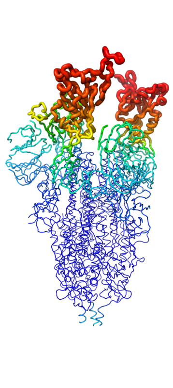
Figure 1. Spike protein shown in "B-Factor"; depicting mobility and flexibility of different portions. Depicted in red are the most mobile, whilst dark blue are the least mobile. The 2 red portions depict RBDs, which correspond to 1-up and 2-up conformational states.
Throughout the entire process, the spike protein has 3 main conformations. An conformation; an active, "open" conformation; and a conformation mentioned previously[3][5][4]. In the closed conformation, the RBDs of each monomer are tucked inwards, preventing interaction. In the open conformation, however, 1 or more of these RBDs can be in the "up" conformation, meaning they are exposed and able to interact within the extracellular space. Mainly, there exits a "" and "" conformation in this phase[5][4]. Depicted in Figure 1, the RBDs of the spike protein have the highest mobility, which further support the many conformational changes in which they are involved. Most of the depictions of the minibinders bound to the spike protein show the spike protein in the 2-up conformation.
ACE2
ACE2 is a carboxypeptidase present on cell surfaces that is responsible for the degradation of angiotensin II. It is a critical enzyme in the suppression of the renin-angiotensin system. This improves both cardiovascular and renal systems, as well as abates acute respiratory distress syndrome (ARDS). It does the 2 former via the RAS System's role in the regulation of blood pressure, renal function, water homeostasis, electrolyte balance, and/or inflammation[6]. The critical role that this enzyme plays in the regulation of this system is what results in the adverse symptomology observed in victims of the SARS-COV-2 virus. The ACE2 receptor is considered the only essential receptor in the SARS-COV-2 viral mechanism, and thus the collateral debilitation of ACE2 results in the adverse respiratory effects including ARDS, pulmonary edema, destruction of alveolar structures, and others[6]. This relationship was further proven when ACE-2 deficient mice had developed these effects at higher rates compared to the wild type[7].
As mentioned previously, all of the S1 subunit domains play important roles in the binding to ACE2. The surface area of the NTD and CTD are particularly important, along with the direct interactions observed in the RBD. Whilst ACE2 is not the focus of this article, understanding its role in the infection pathway of COVID 19, as well as how it binds to the spike protein will assist in understanding the design and functional processes of the minibinders.
Minibinders
Structure
Function
Design
Implications
This is a sample scene created with SAT to by Group, and another to make of the protein. You can make your own scenes on SAT starting from scratch or loading and editing one of these sample scenes.

