From Proteopedia
proteopedia linkproteopedia link AtPIN3
![Figure 1: Auxin (IAAH) enters the cell through influx transporter passes directly through the plasma membrane. Auxin dissociates to release a proton (H+) and anion (IAA-) in the cytoplasm due to higher pH. Due to its charge, it requires PIN proteins to carry it out of the cell. Once it reenters the apoplast it can bind to H again and it moves to the next cell Image by Jen Valenzuela [(CC-BY-NC)]](/wiki/images/thumb/7/74/PIN3.png/250px-PIN3.png)
Figure 1: Auxin (IAAH) enters the cell through influx transporter passes directly through the plasma membrane. Auxin dissociates to release a proton (H+) and anion (IAA-) in the cytoplasm due to higher pH. Due to its charge, it requires PIN proteins to carry it out of the cell. Once it reenters the apoplast it can bind to H again and it moves to the next cell Image by Jen Valenzuela [(CC-BY-NC)]
PIN-FORMED (PIN) proteins in plants are responsible for the polar transport [1] of plant hormone auxin (Figure 1) alongside AUXIN TRANSPORTER PROTEIN 1 (AUX1) and ATP-BINDING CASSETTE (ABC) transporters. The polar transport of Auxin is crucial for proper plant growth and development. There are 8 PIN proteins divided into two subfamilies – six long PINs (PIN1-PIN4, PIN6 and PIN7) and two short PINs (PIN 5 and PIN8) that localize in the plasma membrane and the endoplasmic reticulum respectively. AtPIN3 is a long PIN that shares atleast 54% similarity with other long PINS[1].
Function
Auxin and hence the PIN proteins are involved in many processes like embryogenesis, organogenesis, cell fate determination, and cell division. It also contributes to trophic responses like gravitropism and phototropism.
Mutation of PIN genes or their improper localization may lead to many developmental defects like shorter roots, reduced number of lateral roots, root meristem collapse, defective columella cells, abnormal cotyledons and altered leaf venation
| Structure
AtPIN3 is its is a homodimer with 10 transmembrane (TM1-TM10) domains each (Figure 2). Both the N and the C terminal of the protein lie on the extracellular side. The 10 TM domains are divided into two groups a scaffold domain (TM1–2 and 6–7) and a transport domain (TM3–5 and 8–10).
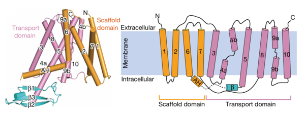 Figure 2:The transmembrane domains and cytoplasmic domains of one chain represented in their proper confirmation and as a simplified diagram. Figure obtained from: Su, N., Zhu, A., Tao, X. et al. Structures and mechanisms of the Arabidopsis auxin transporter PIN3. Nature 609, 616–621 (2022).Figure 1F and 1G The helices 1, 2 and 7 of the scaffold domains are involved in dimerization through symmetric interactions with a surface area of 1516 Å. The tilted TM7 interacts with TM 1, TM2 and TM7 of the other subunit through hydrophobic packaging. TM2 establishes hydrophobic packaging at the base of the dimer. This combined scaffold domain remains static and has the transporter domain on either side of it.
|
Figure 3:A clip taken from the YouTube video entitled: Structure and mechanism of the plant PIN-FORMED auxin transporter Posted by Nanion Technologies Link:
https://www.youtube.com/watch?v=NPQK7T_UhrI Time stamp: 29:57 - 30:31 Speakers: Asc prof. Bjørn Panyella Pedersen. Dept of Molecular Biology and Genetics Aarhus University, Denmark Asc prof. Ulrich Hammes. Plant Systems Biology Technical University of Munich, Germany
The transport domain is predicted to undergo up-down rigid-body motion in an elevator-like model (Figure 3). Two weak helices TM4 and TM9 break in the middle and cross and connect to each other as short loops. These may provide a substrate binding site and allow for confirmational changes during auxin transport. A solvent accessible pathway is present between the scaffold and the transport domain. This was suggested as the location for the . The elevator model is supported by a structural alignment of PIN3 in its apo and IAA bound state, which shows a movement of the transport domain 2-3 Å towards the scaffold domain once IAA binds(Figure4).
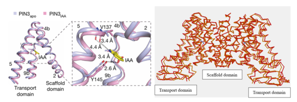
Figure 4:The local confirmation change observed between the structures of PIN3apo and PIN3IAA showing the change of state due to binding of IAA.Figure obtained from: Su, N., Zhu, A., Tao, X. et al. Structures and mechanisms of the Arabidopsis auxin transporter PIN3. Nature 609, 616–621 (2022).Figure 2I and an overall confirmation change observed in the transport domain created using Topmatch a protein structure comparison created by Wiederstein & Sippl (2020) Link:
https://topmatch.services.came.sbg.ac.at/index_jsmol.html?query=7xxb&qname=7xxb&target=7wks&tname=7wksThe protein has a cytosolic domain which contains the cytosolic extension of ,an Amphiphilic helix (AH) and 3 beta strands(β1-3). The loop between the AH and β1-3 has many phosphorylation sites that regulate the subcellular localization and transport activity of the protein.
References
- ↑ Su, N., Zhu, A., Tao, X. et al. Structures and mechanisms of the Arabidopsis auxin transporter PIN3. Nature 609, 616–621 (2022).DOI:https://doi.org/10.1038/s41586-022-05142-w
![Figure 1: Auxin (IAAH) enters the cell through influx transporter passes directly through the plasma membrane. Auxin dissociates to release a proton (H+) and anion (IAA-) in the cytoplasm due to higher pH. Due to its charge, it requires PIN proteins to carry it out of the cell. Once it reenters the apoplast it can bind to H again and it moves to the next cell Image by Jen Valenzuela [(CC-BY-NC)]](/wiki/index.php/Image:PIN3.png)


