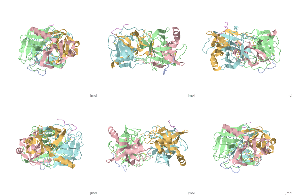
4cha
From Proteopedia
| |||||||||
| 4cha, resolution 1.68Å () | |||||||||
|---|---|---|---|---|---|---|---|---|---|
| Activity: | Chymotrypsin, with EC number 3.4.21.1 | ||||||||
| |||||||||
| |||||||||
| |||||||||
| Resources: | FirstGlance, OCA, RCSB, PDBsum | ||||||||
| Coordinates: | save as pdb, mmCIF, xml | ||||||||
STRUCTURE OF ALPHA-*CHYMOTRYPSIN REFINED AT 1.68 ANGSTROMS RESOLUTION
Overview
Diffraction data for alpha-chymotrypsin crystals at -10 degrees C were measured at 1.68 A resolution and refined by restrained structure-factor least-squares refinement. The two independent chymotrypsin molecules in the crystallographic asymmetric unit were refined independently. The overall structure of alpha-chymotrypsin is little changed from published co-ordinates. The root-mean-square shift of C alpha co-ordinates is 0.42 A, co-ordinates for the two molecules showing a root-mean-square difference of 0.19 A. Certain regions with high disorder (residues 9 to 14, 73 to 79) remain difficult to interpret and several side-chains are disordered. Some water molecule positions have been changed. The absence of the tosyl group has made a significant difference to the refined structure at the active site. This now agrees closely with other enzymes of the trypsin family that have been refined at high resolution. There is a strong hydrogen bond between N epsilon 2 (His57) and O gamma (Ser195) in the free enzyme, in line with the published description of the charge relay system.
About this Structure
4CHA is a Protein complex structure of sequences from Bos taurus. Full crystallographic information is available from OCA.
Reference
Structure of alpha-chymotrypsin refined at 1.68 A resolution., Tsukada H, Blow DM, J Mol Biol. 1985 Aug 20;184(4):703-11. PMID:4046030 Page seeded by OCA on Sun May 4 22:22:07 2008


