This old version of Proteopedia is provided for student assignments while the new version is undergoing repairs. Content and edits done in this old version of Proteopedia after March 1, 2026 will eventually be lost when it is retired in about June of 2026.
Apply for new accounts at the new Proteopedia. Your logins will work in both the old and new versions.
Tedsandbox
From Proteopedia
I think a line at the top looks nice
|
Need more help?
Need more help?
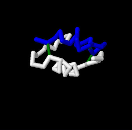
Perhaps it was confusing because you are not used to seeing this protein as a dimer.
Last look.
Click on the disulfide bonds or the white alpha helix for more information.

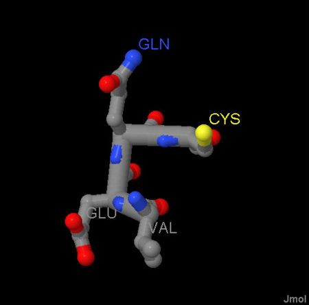
Click on the amino acids for more information.