This old version of Proteopedia is provided for student assignments while the new version is undergoing repairs. Content and edits done in this old version of Proteopedia after March 1, 2026 will eventually be lost when it is retired in about June of 2026.
Apply for new accounts at the new Proteopedia. Your logins will work in both the old and new versions.
User:Eleanor Crabb/Sandbox 1
From Proteopedia
|
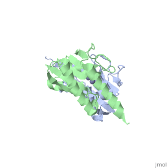
The interactive image on the right shows the protein with the pdb code 1z3a. You can rotate the molecule using your mouse and zoom by holding down the shift key while you move the mouse.
You will note that in this protein, there are two chains shown in green and blue. Let us concentrate on just a , chain A. To see this click on the link in green.
This is a cartoon representation of the protein, showing a schematic representation of the back bone of the protein. You may be more familiar with the following molecular representations (note that the H atoms are not shown):
- - this gives an indication of the size and shape of the protein
Let us now look at just a single length of the chain.
If we now draw a line through the backbone of the chain Let us return to the whole chain. The arrangement of the residues is the primary structure - coloured according to . Secondary structure
