Gamma Carbonic Anhydrase

From Proteopedia

Jump to: navigation, search

Contents

Gamma Carbonic Anhydrase

Introduction/Background

Gamma carbonic anhydrase is an enzyme that is produced by the methanoarchaeon Methanosarcina thermophila [1]. Gamma carbonic anhydrase, often referred to as Cam (for carbonic anhydrase of M. thermophila) is part of a class of enzymes known as the carbonic anhydrases, which catalyze the reversible hydration of carbon dioxide (CO2) to the bicarbonate ion (HCO3-). There are three distinct classes of carbonic anhydrases, the alpha (α) form found in mammals, the beta (β) form found in bacteria and higher order photosynthetic plants, and the gamma (γ) form found in archaea. Evolutionary studies show that the three classes of carbonic anhydrases do not have common ancestry, however additional studies into the catalytic mechanism of γ-carbonic anhydrase indicate that α and γ carbonic anhydrases similar use of a zinc hydroxide mechanism to oxidize CO2.[2]


Function of Gamma Carbonic Anhydrase

The main biological function of γ-carbonic anhydrase in M. thermophila is to catalyze the hydration of CO2 to HCO3- ion, in order to reduce the concentration of CO2 produced in acetate metabolism. M. thermophila are methanogenic anaerobes which produce methane by reducing the methyl groups of a variety of organic molecules such as acetate, methanol and methylamines [3]. When high concentrations of acetate are available, M. thermophila is able to switch its metabolism to use acetate to produce methane from reduced the methyl group and carbon dioxide from the oxidized carbonyl group [1] [3]. The role of γ-carbonic anhydrase in acetate metabolism is drive forward the reaction of acetate to methane and CO2 by removing the CO2 concentration by converting it to HCO3- outside the cell [1] [3].


Structure

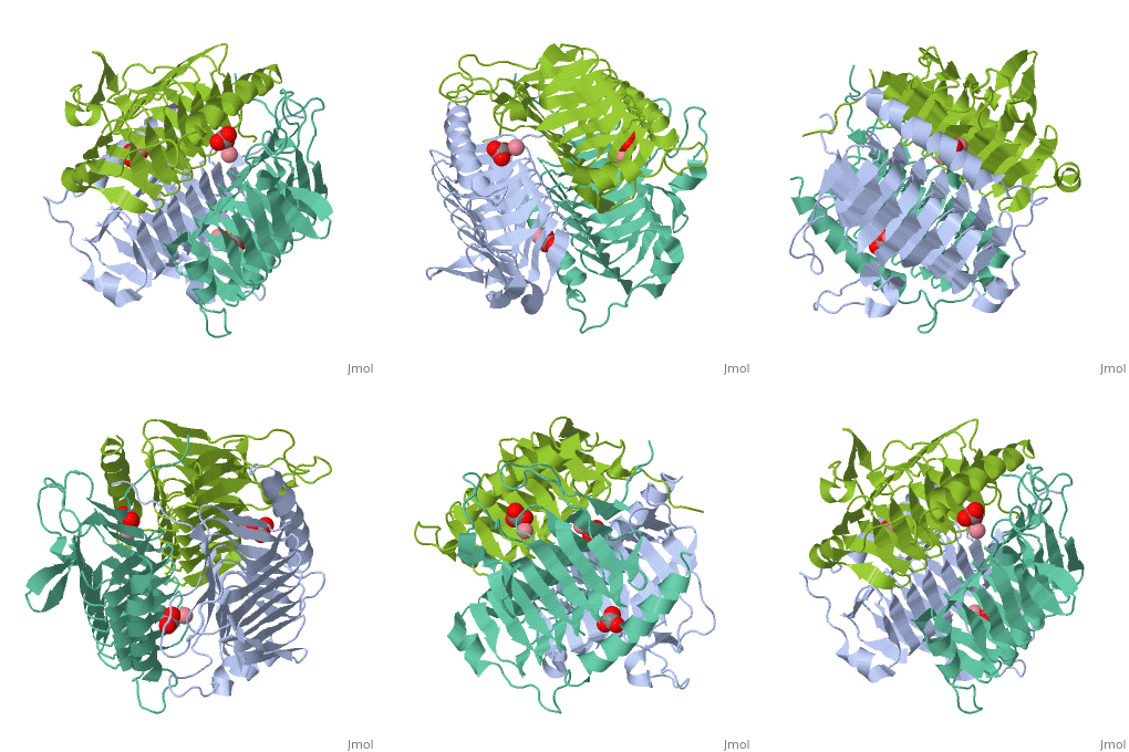
According to the Structural Classification of Proteins (SCOP), γ-carbonic anhydrase is part of the Trimeric LpxA Enzyme superfamily, which is characterized by single-stranded polypeptides with left-handed beta-helix fold. The overall structure of γ-carbonic anhydrase is a trimer complex composed of left-handed beta-helix monomers. The beta-helix consists of three untwisted, parallel sheets that are connected by left-handed crossovers [1]. Like all the carbonic anhydrase proteins, γ-carbonic anhydrase contains a metal ion ligand in its active site. The unique feature of γ-carbonic anhydrase is its ability to utilize two metal ions equivalently for its active site, the zinc ion, Zn2+ and the cobalt ion, Co2+. Studies have shown that the enzyme is able to use these two ions interchangeable depending on their availability [2]. While studies indicate that there are no significant differences between zinc-bound and cobalt-bound γ-carbonic anhydrase structures, the catalytic mechanism for binding of carbon dioxide is different between the three classes of carbonic anhydrases. In both zinc-bound and cobalt-bound γ-carbonic anhydrases there are three histidine residues (His81, His117 and His122 residues) that coordinate the ion with the active site [1]. In addition to the three histidines, there are several other residues which have been identified to play an important role in the active site catalytic mechanism. For example the glutamine residue Gln75 and the asparagine residue Asn73 have been found to help orient the cobalt ion for attack on the carbon dioxide, and the asparagine residue Asn202 prepares the carbon dioxide, by polarization, for attack by the cobalt ion [4].

PDB ID 1qre

Drag the structure with the mouse to rotate

Catalytic Mechanism

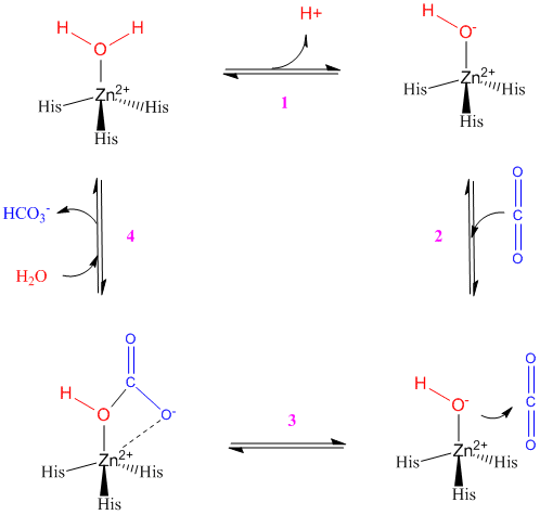
The catalytic mechanism for γ-carbonic anhydrase active site is shown below, using zinc as the metal ion ligand. In the case of cobalt as the metal ion ligand, there is an additional water molecule present in the active site. In the first step the zinc ion is coordinated with two water molecules (only one is shown on the diagram) and the three histidine residues. The Glu62 residue donates electrons to one of the water molecules, which in turn donates the electrons to the neighbouring water molecule (this is shown by the hydrogen ion on the diagram).The result is a zinc hydroxide. In the next step (2 to 3) the zinc hydroxide attacks the incoming carbon dioxide molecule, which is held in place in the proper orientation by Asn202 and Gln75 residues (not shown), to form the bicarbonate ion and displace one of the water molecules. In the final steps (3 to 4), the bicarbonate ion is displaced through hydrogen bonding with the Glu62 residue and the addition of two water molecules to the zinc ion. The water molecules replace the two oxygen molecules that were bound to the zinc ion, thus regenerating the enzyme [1] [4].


Image:Carbonic Anhydrase Mechanism.gif


3D structues of γ-carbonic anhydrase

Carbonic anhydrase

References

  1. 1.0 1.1 1.2 1.3 1.4 1.5 Iverson TM, Alber BE, Kisker C, Ferry JG, Rees DC. A closer look at the active site of gamma-class carbonic anhydrases: high-resolution crystallographic studies of the carbonic anhydrase from Methanosarcina thermophila. Biochemistry. 2000 Aug 8;39(31):9222-31. PMID:10924115
  2. 2.0 2.1 Alber BE, Colangelo CM, Dong J, Stalhandske CM, Baird TT, Tu C, Fierke CA, Silverman DN, Scott RA, Ferry JG. Kinetic and spectroscopic characterization of the gamma-carbonic anhydrase from the methanoarchaeon Methanosarcina thermophila. Biochemistry. 1999 Oct 5;38(40):13119-28. PMID:10529183
  3. 3.0 3.1 3.2 Alber BE, Ferry JG. A carbonic anhydrase from the archaeon Methanosarcina thermophila. Proc Natl Acad Sci U S A. 1994 Jul 19;91(15):6909-13. PMID:8041719
  4. 4.0 4.1 Zimmerman SA, Ferry JG. Proposal for a hydrogen bond network in the active site of the prototypic gamma-class carbonic anhydrase. Biochemistry. 2006 Apr 25;45(16):5149-57. PMID:16618104 doi:10.1021/bi052507y
Proteopedia Page Contributors and Editors (what is this?)

Michal Harel, Meagan Moi, Alexander Berchansky

Personal tools