Journal:Acta Cryst F:S2053230X21013455

From Proteopedia

Jump to: navigation, search

Crystal structure of betaine aldehyde dehydrogenase from Burkholderia pseudomallei

Dylan K. Beard, Sandhya Subramanian, Jan Abendroth, Thomas E. Edwards, Peter J. Myler, and Oluwatoyin A. Asojo [1]


Molecular Tour
Burkholderia pseudomallei is a rod-shaped, motile, flagellated, soil-dwelling gram-negative proteobacterium of the Burkholderiaceae family that thrives in tropical and subtropical regions. B. pseudomallei causes melioidosis, a deadly emerging opportunistic infection mainly of the immunocompromised. B. pseudomallei is transmitted through open wounds, contact with contaminated soil and water, ingestion, or inhalation, and is also a potential biological warfare agent. The Seattle Structural Genomics Center for Infectious Disease (SSGCID) has determined and solved X-ray structures of a potential drug target betaine aldehyde dehydrogenase (BADH) from B. pseudomallei (BpBADH):

  • Monomer of apo BpBADH, colored in rainbow from blue (N-terminus) to red (C-terminus); 6wsa.
  • Dimer of BpBADH with NAD, monomers are shown as green and cyan ribbons, with NAD shown as ball-and-sticks; 6wsb.

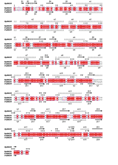
BpBADH has a prototypical BADH topology and shares considerable structure and sequence similarity with the ortholog from P. aeruginosa (PaBADH); see static image below:

Figure 2 Structural and primary sequence alignment of BpBADH and PaBADH. The secondary structure elements shown are alpha helices (α), 310-helices (η), beta strands (β), and beta turns (TT). Identical residues are shown in white on a red background and conserved residues in red.

The structures are similar to those of BADH from Pseudomonas aeruginosa (PaBADH). The co-factor binding domains of BpBADH (6wsb) and PaBADH (4caz) are well conserved (identical residues of both structures are labeled in green, while non-identical in red):

  • Co-factor binding domain of BpBADH (6wsb).
  • Co-factor binding domain of PaBADH (4caz).
  • Superposition of BpBADH and PaBADH (BpBADH is colored in green, PaBADH is in olive).

PaBADH is inhibited by the drug disulfiram which is an approved drug. Both structures show the conserved catalytic cysteine irreversibly inhibited by disulfiram. Our preliminary analysis could facilitate drug repurposing studies for melioidosis. This project is an educational collaboration between the SSGCID and Hampton University. The SSGCID consortium is directed by Dr. Peter Myler (principal investigator) and comprises many different scientists working at multiple centers towards determining the three-dimensional structures of proteins from biodefense organisms and emerging infectious diseases. Dylan K. Beard was part of a pilot Hampton University Chemistry Education and Mentorship Course-based undergraduate research (HU-ChEM CURES) funded by the NIGMS.

PDB references: betaine aldehyde dehydrogenase, 6wsa; bound to cofactor, 6wsb.

References

  1. Beard DK, Subramanian S, Abendroth J, Dranow DM, Edwards TE, Myler PJ, Asojo OA. Crystal structure of betaine aldehyde dehydrogenase from Burkholderia pseudomallei. Acta Crystallogr F Struct Biol Commun. 2022 Feb 1;78(Pt 2):45-51. doi:, 10.1107/S2053230X21013455. Epub 2022 Jan 27. PMID:35102892 doi:http://dx.doi.org/10.1107/S2053230X21013455
Drag the structure with the mouse to rotate

Proteopedia Page Contributors and Editors (what is this?)

Alexander Berchansky, Jaime Prilusky

This page complements a publication in scientific journals and is one of the Proteopedia's Interactive 3D Complement pages. For aditional details please see I3DC.
Personal tools