User:Camryn Jiles/Sandbox 1
From Proteopedia
Neuropeptide: Orexin
This is a default text for your page Camryn Jiles/Sandbox 1. Click above on edit this page to modify. Be careful with the < and > signs. You may include any references to papers as in: the use of JSmol in Proteopedia [1] or to the article describing Jmol [2] to the rescue.
IntroductionOrexin, also known as hypocretin, are a group of neuropeptides identified in the late 1990’s, located in the lateral region of the hypothalamus. Neuropeptides are short strings of amino acids that are synthesized and released by neurons in the brain. These chemical messengers will typically bind to G protein-coupled receptors (GPCRs) to regulate neural activity. Human neuropeptide Y consists of three ligands (NPY, PYY and PP) and four receptors (Y1R, Y2R, Y3R, Y4R and Y5R). NPY is abundant in the brain and its structure consists of a C-terminal segment that forms an α-helix; its N terminal is flexible and relatively unstructured. The receptors act as an antagonist, when the ligand and receptor come together forming a complex, resulting in an extracellular conformational change. The C terminus of NPY will penetrate the transmembrane core of the receptor which in turn, activates the G protein-coupled receptor. One key characteristic that is indicative of this type of activated GPCR includes interaction “between R138 of the (D/E)R(Y/H) motif and Y231 and Y320 of the NPY motif” (Park 2).
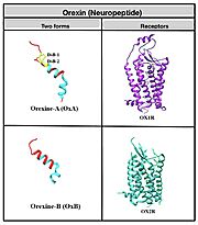
Function & StructureOrexin's primarily play a role in the central nervous system and are responsible for arousal, wakefulness and appetite. Additionally, they are capable of various physiology functions, regulating reproductive and neuroendocrine functions, gastrointestinal motility, blood pressure, metabolism and energy balance. Orexin consists of 131 amino acids and is broken down into two forms: orexin-A (OxA) which consists of 33 amino acids and orexin-B (OxB), consisting of 28 amino acids. OxA consists of a pyroglutamyl residue on the N terminus, two disulfide bridges and an amidated C-terminus. OxB also contains an amidated C-terminus. Both proteins also contain two ɑ-helices within their structure. OxA and OxB activates orexin receptor type 1(OX1R) and orexin receptor type 2 (OXR2) that act on G protein-coupled receptors located on effector cells. It is worth noting that OX1R has a better affinity for OxA and OX2R for OxB. This signaling pathway and resulting influx of calcium into the intracellular space, is what allows orexin to mediate the various biological actions across the central and peripheral nervous system. RelevanceExpression of orexin receptors isn’t just limited to the brain. They can be found in the adrenal glands, pancreas, vertebrate testis, placenta, kidneys and more. Stress is a major stimulator for increased orexin production, as it induces the release of cortisol from the adrenals. The relationship between orexin and stress is inversely proportional, and regulation of acute stress is better understood then long term stress; “orexin promote the acute behavioral and neuroendocrine response to stress, and acute stress activates orexin neurons” (Grafe). Glucose is another stimulator of orexin release and comes from the beta cells of the pancreas. Whenever the body is hungry, the plasma concentration of orexin increases which subsequently increases appetite. Though the entire process of how this works is still misunderstood, it is suggested that ghrelin (hunger hormone) activates orexin neurons independent of NPY. There is also a relationship between obesity and orexin worth briefly noting. A recent study showed that orexin increases adipocyte glucose uptake and stimulates adiponectin release in rats. Additionally, lipoprotein lipase in adipocytes increases when exposed to orexin. In conclusion, this study showed that orexin increases the enzyme that degrades fat hence, removal of orexin causes obesity due to decreased energy and browning of fat (Adeghate 6). GPCR’s are the largest, diverse family of human transmembrane proteins and play an important role in cell signaling. This factor makes them ideal for drug targets to achieve numerous therapeutic effects; with this in mind it is important to get a broad understanding of the structure and binding dynamics of GPCRs. OX1R and OX2R are class A GPCRs which is the largest of the four classes, and most diverse in humans. Its structure consists of a “seven-transmembrane helices domain, three extracellular loops and three intracellular loops with ligand binding pockets” (Vu 1). Peptide hormones like neuropeptides, though very diverse, undergo similar processing to get to their active form; many undergo different post-translational modifications to achieve this by inhibiting exopeptidase, bromination, lipidation, disulfide bridge formation or differential proteolysis. Neuropeptides specifically undergo C-terminal amidation which acts to reduce the charge of the peptide which improves potency, metabolic stability and increases ability to resist enzymatic degradation. InhibitionDue to orexins' strong regulation on sleep, decreased production can lead to narcolepsy. Narcolepsy is a chronic sleep disorder characterized by overwhelming drowsiness and sudden attacks of sleep. In addition, dysregulation can lead to extreme happiness or extreme sadness due to orexin's effect on mood. Abnormal expression of orexin receptor type 1 can cause cancer and the development of metabolic diseases, like diabetes, can also occur. However, GPCR peptide drugs can aid in reducing these symptoms by modulating the orexin signaling pathway– ultimately inhibiting it. Antagonistic drugs called orexin-receptor antagonists (SORAs) have been made to target orexin receptors, consisting of the antagonist compound suvorexant. Upon binding, suvorexant adopts a π-stacked conformation and binds deep to the active site of the receptor and prevents transmembrane helix activation. A molecular binding simulation between OxB and OX1R showed that “mutation in the alanine residue of K120, P123, Y124, N318, F340, T341, H344 and W345 located in the TM2, TM3, TM6 and TM7 reduces binding affinity and inactivates the intracellular release of calcium ions.
ConclusionIn conclusion, orexin and its receptors play a major role in maintaining overall homeostasis within the body. Its involvement in various neural activity, including other organ systems makes it an important biochemical protein to obtain feeding behaviors, energy balance, the sleep-wake cycle and more. One future area of study that may be interesting to explore would be further understanding of the relationship between orexin and long term stress, as it is misunderstood at the present time.
ReferencesAdeghate, E., Lotfy, M., D’Souza, C., Alseiari, S. M., Alsaadi, A. A., & Qahtan, S. A. (2020). Hypocretin/orexin modulates body weight and the metabolism of glucose and insulin. Diabetes/Metabolism Research and Reviews, 36(3), e3229. https://doi-org.proxy.library.maryville.edu/10.1002/dmrr.3229 Alain, C., Pascal, N., Valérie, G., & Thierry, V. (2021). Orexins/Hypocretins and Cancer: A Neuropeptide as Emerging Target. Molecules (Basel, Switzerland), 26(16). https://doi.org/10.3390/molecules26164849 Grafe, L. A., & Bhatnagar, S. (2018). Orexins and stress. Frontiers in neuroendocrinology, 51, 132–145. https://doi.org/10.1016/j.yfrne.2018.06.003 Hong, C., Byrne, N. J., Zamlynny, B., Tummala, S., Xiao, L., Shipman, J. M., Partridge, A. T., Minnick, C., Breslin, M. J., Rudd, M. T., Stachel, S. J., Rada, V. L., Kern, J. C., Armacost, K. A., Hollingsworth, S. A., O’Brien, J. A., Hall, D. L., McDonald, T. P., Strickland, C., … Hollenstein, K. (2021). Structures of active-state orexin receptor 2 rationalize peptide and small-molecule agonist recognition and receptor activation. Nature Communications, 12(1), 815. https://doi-org.proxy.library.maryville.edu/10.1038/s41467-021-21087-6 Li, S.-B., Jones, J. R., & de Lecea, L. (2016). Hypocretins, Neural Systems, Physiology, and Psychiatric Disorders. Current Psychiatry Reports, 18(1), 7. https://doi.org/10.1007/s11920-015-0639-0 Park, Chaehee, et al. “Structural Basis of Neuropeptide Y Signaling through Y1 Receptor.” Nature Communications, vol. 13, no. 1, Feb. 2022, p. 853. EBSCOhost, https://doi-org.proxy.library.maryville.edu/10.1038/s41467-022-28510-6. Vu, O., Bender, B. J., Pankewitz, L., Huster, D., Beck-Sickinger, A. G., & Meiler, J. (2021). The Structural Basis of Peptide Binding at Class A G Protein-Coupled Receptors. Molecules (Basel, Switzerland), 27(1). https://doi-org.proxy.library.maryville.edu/10.3390/molecules27010210
| ||||||||||||

