Sandbox Reserved 459

From Proteopedia

Jump to: navigation, search
This Sandbox is Reserved from 13/03/2012, through 01/06/2012 for use in the course "Proteins and Molecular Mechanisms" taught by Robert B. Rose at the North Carolina State University, Raleigh, NC USA. This reservation includes Sandbox Reserved 451 through Sandbox Reserved 500.
To get started:
  • Click the edit this page tab at the top. Save the page after each step, then edit it again.
  • Click the 3D button (when editing, above the wikitext box) to insert Jmol.
  • show the Scene authoring tools, create a molecular scene, and save it. Copy the green link into the page.
  • Add a description of your scene. Use the buttons above the wikitext box for bold, italics, links, headlines, etc.

More help: Help:Editing

For more help, look at this link: http://www.proteopedia.org/wiki/index.php/Help:Getting_Started_in_Proteopedia

Contents

Amylase


Introduction


Amylase is categorized in a class of enzymes known as hydrolases. Hydrolases are enzymes which use water to cleave chemical bonds, usually dividing a large molecule into two smaller molecules. Examples of common hydrolases include esterases, proteases, glycosidases, nucleosidases, and lipases. Hydrolases carry out important degradative reactions in the body. During digestion, lipases hydrolyze lipids and proteases convert protein to amino acids. Hydrolases cleave large molecules into fragments used for synthesis , the excretion of waste materials, or as sources of carbon for the production of energy. [1] Amylase is an enzyme that catalyses the breakdown of starch into sugars. Amylase is present in human saliva, where it begins the chemical process of digestion. Food that contains much starch but little sugar, such as rice and potato, taste slightly sweet as they are chewed because amylase turns some of their starch into sugar in the mouth. The pancreas also makes amylase (alpha amylase) to hydrolyse dietary starch into disaccharides and trisaccharides which are converted by other enzymes to glucose to supply the body with energy. Plants and some bacteria also produce amylase. As diastase, amylase was the first enzyme to be discovered and isolated. Amylase in all organisms has the function of breaking down complex carbohydrates.

Structure of human salivary alpha-amylase

Drag the structure with the mouse to rotate


Structure


To examine the crystal structure of human salivary amylase, X-ray crystallography was used with a resolution of 1.60 Å. [2] The active site of alpha-amylase contains a trio of acidic groups that do most of the work. Ca2+ is a common for alpha amylase.The Ca2+ ion is bound to Asnl00, Arg158, Asp167, His201 and three water molecules. The Cl- ion is bound to Arg195, Asn298 and Arg337 and one water molecule. The highly mobile glycine-rich loop 304-310 may act as a gateway for substrate binding and be involved in a `trap-release' mechanism in the hydrolysis of substrates. Strategic placement of calcium and chloride ions, as well as histidine and tryptophan residues may play a role in differentiating between the glycone and aglycone ends of the polysaccharide substrates. Salivary amylase also possesses a suitable site for binding to enamel surfaces and provides potential sites for the binding of bacterial adhesins. . Salivary amylase folds into a multidomain structure consisting of three domains, A, B and C. Domain A has a (a/b)8- barrel structure, domain B has no definite topology and domain C has a Greek-key barrel structure. Circular dichroism spectroscopic data revealed the native alpha-amylase to contain 25% , 21% , and 54% random coils. [3] It is generally assumed that in proteins hydrophobic residues are not favorable at solvent-exposed sites, and that amino acid substitutions on the surface have little effect on protein thermostability. Contrary to these assumptions, hyperthermostable variants of alpha amylase have been identified that result from the incorporation of at the surface. [4]


Mechanism of Action


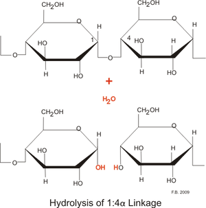
Hydrolysis of sugar using Amylase

Hydrolysis of starch or oligosaccharides by mammalian amylases, in general, results in maltose as the leaving group. The active site of these amylases harbors three aromatic residues Trp59, Tyr62, and Tyr151, which provide stacking interactions to the bound glucose moieties. The hydrolysis is catalyzed by three carboxyl groups and its starts from a water nucleophilic attack and opening of the glucose ring in the catalytic center rather than from protonation of the glycosidic oxygen. One of the carboxylic acids in the active site acts as the catalytic nucleophile during the formation of the intermediate. A second carboxylic acid operates as the acid/base catalyst, supporting the stabilization of the transition states during the hydrolysis. The chain length of the oligosaccharide has an impact on both the rate of the hydrolytic degradation and the product pattern produced by a-amylase. Oligomers with more than six glucose units are hydrolyzed more rapidly, because they are more akin to the natural substrates of a-amylase. [5]


Medical Implications or Possible Applications

  1. 1. Blood serum amylase- Blood serum amylase may be measured for purposes of medical diagnosis. A normal concentration is in the range 21-101 U/L. A higher than normal concentration may reflect one of several medical conditions, including acute inflammation of the pancreas. When the pancreas is diseased or inflamed, amylase is released into the blood. Blood serum amylase can also aid in diagnosing a perforated peptic ulcer, torsion of an ovarian cyst, strangulation ileus, macroamylasemia and mumps. Amylase may be measured in other body fluids, including urine and peritoneal fluid.
  1. 2. Amylase deficiency- One of the leading cause for amylase deficiency is a high carbohydrate diet. High volume of complex carbohydrate in the diet results in increased demand for amylase to convert them into simple sugars. After prolonged period of time, the levels of amylase fall below normal which result in amylase deficiency. Amylase is involved in anti-inflammatory reactions such as those caused by the release of histamine and similar substances. The inflammatory response usually occurs in organs which are in contact with the outside world such as the lungs and skin. Therefore, an amylase deficiency can include skin problems such as psoriasis, eczema, hives, insect bites, allergic bee and bug stings, atopic dermatitis, and all types of herpes. Lung problems, including asthma and emphysema, require amylase plus other enzyme formulas depending on the particular condition. [6]

Amylase Inhibitors


Amylase inhibitors are also known as starch blockers because they contain substances that prevent dietary starches from being absorbed by the body.

  1. Nonproteinaceous inhibitors- The class of nonproteinaceous inhibitors contains diverse types of organic compounds such as acarbose, isoacarbose, acarviosine-glucose, hibiscus acid and the cyclodextrins. The two hibiscus acid forms, purified from Roselle tea (Hibiscus sabdariffa), the acarviosine-glucose, the isoacarbose and a-, b- and c-cyclodextrins are highly active against porcine and human pancreatic a-amylase. The inhibitory activity of these compounds against a-amylases is due in part to their cyclic structures, which resemble a-amylase substrates and therefore bind to a-amylase catalytic sites.
  2. Cereal-type a-amylase inhibitors- a-Amylase inhibitors of the cereal family are composed of 120-160 amino-acid residues forming five disulfide bonds. These inhibitors are also known as sensitizing agents in humans upon repeated exposure, causing allergy, dermatitis and baker's asthma associated with cereal flour. The bifunctional a-amylase/trypsin inhibitor is another prototype of the cereal inhibitor family. This inhibitor is a stable monomer of 122 amino acids with five disulfide bonds, which is resistant to urea, guanidine hydrochloride and thermal denaturation. [7]

References

  1. Gibbs, J. (2003). Hydrolase. Retrieved from http://www.chemistryexplained.com/Ge-Hy/Hydrolase.html
  2. PDB file. Ramasubbu, N. (1996). Structure of human salivary alpha-amylase at 1.6 a resolution: implications for its role in the oral cavity. . [Web Graphic]. Retrieved from http://www.rcsb.org/pdb/explore/explore.do?structureId=1SMD
  3. Rao , U. (2008). Biophysical and biochemical characterization of a hyperthermostable and ca2 -independent alpha-amylase of an extreme thermophile geobacillus thermoleovorans. Applied Biochemistry and Biotechnology, 150(2), 205-219. Retrieved from http://www.ncbi.nlm.nih.gov/pubmed/18443744
  4. Machius, M. (2003). Kinetic stabilization of bacillus licheniformis a-amylase through introduction of hydrophobic residues at the surface. The Journal of Biological Chemistry, 278(13), 11546–11553. Retrieved from http://www.jbc.org/content/278/13/11546.full.pdf
  5. Mishra, P. (2002). The mechanism of salivary amylase hydrolysis: role of residues at subsite s2'. Biochem Biophys Res Commun, 292(2), 468-473. Retrieved from http://www.ncbi.nlm.nih.gov/pubmed/11906186
  6. Symptoms of enzyme deficiencies. (2012). Retrieved from http://enzymes.subtleenergysolutions.com/enzyme-deficiency.html
  7. Franco, O. (2002). Plant a-amylase inhibitors and their interaction with insect a-amylases. European Journal of Biochemistry, 269, 397-412. Retrieved from http://onlinelibrary.wiley.com/doi/10.1046/j.0014-2956.2001.02656.x/pdf
Personal tools