Structural highlights
Function
CASP1_HUMAN Thiol protease that cleaves IL-1 beta between an Asp and an Ala, releasing the mature cytokine which is involved in a variety of inflammatory processes. Important for defense against pathogens. Cleaves and activates sterol regulatory element binding proteins (SREBPs). Can also promote apoptosis.[1] [2]
Evolutionary Conservation
Check, as determined by ConSurfDB. You may read the explanation of the method and the full data available from ConSurf.
Publication Abstract from PubMed
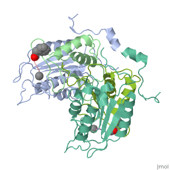
Structural studies of caspase-1 reveal that the dimeric thiol protease can exist in two states: in an on-state, when the active site is occupied, or in an off-state, when the active site is empty or when the enzyme is bound by a synthetic allosteric ligand at the dimer interface approximately 15 A from the active site. A network of 21 hydrogen bonds from nine side chains connecting the active and allosteric sites change partners when going between the on-state and the off-state. Alanine-scanning mutagenesis of these nine side chains shows that only two of them-Arg286 and Glu390, which form a salt bridge-have major effects, causing 100- to 200-fold reductions in catalytic efficiency (k(cat)/K(m)). Two neighbors, Ser332 and Ser339, have minor effects, causing 4- to 7-fold reductions. A more detailed mutational analysis reveals that the enzyme is especially sensitive to substitutions of the salt bridge: even a homologous R286K substitution causes a 150-fold reduction in k(cat)/K(m). X-ray crystal structures of these variants suggest the importance of both the salt bridge interaction and the coordination of solvent water molecules near the allosteric binding pocket. Thus, only a small subset of side chains from the larger hydrogen bonding network is critical for activity. These form a contiguous set of interactions that run from one active site through the allosteric site at the dimer interface and onto the second active site. This subset constitutes a functional allosteric circuit or "hot wire" that promotes site-to-site coupling.
An allosteric circuit in caspase-1.,Datta D, Scheer JM, Romanowski MJ, Wells JA J Mol Biol. 2008 Sep 19;381(5):1157-67. Epub 2008 Jun 20. PMID:18590738[3]
From MEDLINE®/PubMed®, a database of the U.S. National Library of Medicine.
See Also
References
- ↑ Alnemri ES, Fernandes-Alnemri T, Litwack G. Cloning and expression of four novel isoforms of human interleukin-1 beta converting enzyme with different apoptotic activities. J Biol Chem. 1995 Mar 3;270(9):4312-7. PMID:7876192
- ↑ Feng Q, Li P, Leung PC, Auersperg N. Caspase-1zeta, a new splice variant of the caspase-1 gene. Genomics. 2004 Sep;84(3):587-91. PMID:15498465 doi:http://dx.doi.org/S0888-7543(04)00161-2
- ↑ Datta D, Scheer JM, Romanowski MJ, Wells JA. An allosteric circuit in caspase-1. J Mol Biol. 2008 Sep 19;381(5):1157-67. Epub 2008 Jun 20. PMID:18590738 doi:10.1016/j.jmb.2008.06.040