2kdh
From Proteopedia
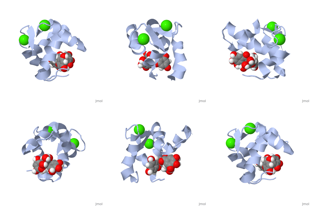
The Solution Structure of Human Cardiac Troponin C in complex with the Green Tea Polyphenol; (-)-epigallocatechin-3-gallate
Structural highlights
DiseaseTNNC1_HUMAN Defects in TNNC1 are the cause of cardiomyopathy dilated type 1Z (CMD1Z) [MIM:611879. Dilated cardiomyopathy is a disorder characterized by ventricular dilation and impaired systolic function, resulting in congestive heart failure and arrhythmia. Patients are at risk of premature death.[1] Defects in TNNC1 are the cause of familial hypertrophic cardiomyopathy type 13 (CMH13) [MIM:613243. A hereditary heart disorder characterized by ventricular hypertrophy, which is usually asymmetric and often involves the interventricular septum. The symptoms include dyspnea, syncope, collapse, palpitations, and chest pain. They can be readily provoked by exercise. The disorder has inter- and intrafamilial variability ranging from benign to malignant forms with high risk of cardiac failure and sudden cardiac death.[2] [3] [4] [5] FunctionTNNC1_HUMAN Troponin is the central regulatory protein of striated muscle contraction. Tn consists of three components: Tn-I which is the inhibitor of actomyosin ATPase, Tn-T which contains the binding site for tropomyosin and Tn-C. The binding of calcium to Tn-C abolishes the inhibitory action of Tn on actin filaments. Evolutionary ConservationCheck, as determined by ConSurfDB. You may read the explanation of the method and the full data available from ConSurf. Publication Abstract from PubMedHeart muscle contraction is regulated by Ca(2+) binding to the thin filament protein troponin C. In cardiovascular disease, the myofilament response to Ca(2+) is often altered. Compounds that rectify this perturbation are of considerable interest as therapeutics. Plant flavonoids have been found to provide protection against a variety of human illnesses such as cancer, infection, and heart disease. (-)-Epigallocatechin gallate (EGCg), the prevalent flavonoid in green tea, modulates force generation in isolated guinea pig hearts (Hotta, Y., Huang, L., Muto, T., Yajima, M., Miyazeki, K., Ishikawa, N., Fukuzawa, Y., Wakida, Y., Tushima, H., Ando, H., and Nonogaki, T. (2006) Eur. J. Pharmacol. 552, 123-130) and in skinned cardiac muscle fibers (Liou, Y. M., Kuo, S. C., and Hsieh, S. R. (2008) Pflugers Arch. 456, 787-800; and Tadano, N., Yumoto, F., Tanokura, M., Ohtsuki, I., and Morimoto, S. (2005) Biophys. J. 88, 314a). In this study we describe the solution structure of the Ca(2+)-saturated C-terminal domain of troponin C in complex with EGCg. Moreover, we show that EGCg forms a ternary complex with the C-terminal domain of troponin C and the anchoring region of troponin I. The structural evidence indicates that the binding site of EGCg on the C-terminal domain of troponin C is in the hydrophobic pocket in the absence of troponin I, akin to EMD 57033. Based on chemical shift mapping, the binding of EGCg to the C-terminal domain of troponin C in the presence of troponin I may be to a new site formed by the troponin C.troponin I complex. This interaction of EGCg with the C-terminal domain of troponin C.troponin I complex has not been shown with other cardiotonic molecules and illustrates the potential mechanism by which EGCg modulates heart contraction. Solution structure of human cardiac troponin C in complex with the green tea polyphenol, (-)-epigallocatechin 3-gallate.,Robertson IM, Li MX, Sykes BD J Biol Chem. 2009 Aug 21;284(34):23012-23. Epub 2009 Jun 20. PMID:19542563[6] From MEDLINE®/PubMed®, a database of the U.S. National Library of Medicine. See AlsoReferences
| ||||||||||||||||||||


