Structural highlights
Function
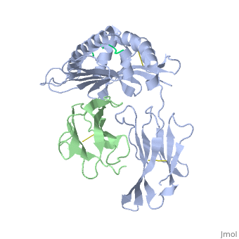
[HA1B_MOUSE] Involved in the presentation of foreign antigens to the immune system. [NCAP_SENDE] Encapsidates the genome in a ratio of one N per six ribonucleotides, protecting it from nucleases. The nucleocapsid (NC) has a helical structure with 13.07 N per turn. The encapsidated genomic RNA is termed the NC and serves as template for transcription and replication. Replication is dependent on intracellular concentration of newly synthesized N, termed N(0), which corresponds to the protein not associated with RNA. In contrast, when associated with RNA, it is termed N. During replication, encapsidation by N(0) is coupled to RNA synthesis and all replicative products are resistant to nucleases. [B2MG_MOUSE] Component of the class I major histocompatibility complex (MHC). Involved in the presentation of peptide antigens to the immune system.
Evolutionary Conservation
Check, as determined by ConSurfDB. You may read the explanation of the method and the full data available from ConSurf.
Publication Abstract from PubMed
The x-ray structures of a murine MHC class I molecule (H-2Kb) were determined in complex with two different viral peptides, derived from the vesicular stomatitis virus nucleoprotein (52-59), VSV-8, and the Sendai virus nucleoprotein (324-332), SEV-9. The H-2Kb complexes were refined at 2.3 A for VSV-8 and 2.5 A for SEV-9. The structure of H-2Kb exhibits a high degree of similarity with human HLA class I, although the individual domains can have slightly altered dispositions. Both peptides bind in extended conformations with most of their surfaces buried in the H-2Kb binding groove. The nonamer peptide maintains the same amino- and carboxyl-terminal interactions as the octamer primarily by the insertion of a bulge in the center of an otherwise beta conformation. Most of the specific interactions are between side-chain atoms of H-2Kb and main-chain atoms of peptide. This binding scheme accounts in large part for the enormous diversity of peptide sequences that bind with high affinity to class I molecules. Small but significant conformational changes in H-2Kb are associated with peptide binding, and these synergistic movements may be an integral part of the T cell receptor recognition process.
Crystal structures of two viral peptides in complex with murine MHC class I H-2Kb.,Fremont DH, Matsumura M, Stura EA, Peterson PA, Wilson IA Science. 1992 Aug 14;257(5072):919-27. PMID:1323877[1]
From MEDLINE®/PubMed®, a database of the U.S. National Library of Medicine.
See Also
References
- ↑ Fremont DH, Matsumura M, Stura EA, Peterson PA, Wilson IA. Crystal structures of two viral peptides in complex with murine MHC class I H-2Kb. Science. 1992 Aug 14;257(5072):919-27. PMID:1323877