This old version of Proteopedia is provided for student assignments while the new version is undergoing repairs. Content and edits done in this old version of Proteopedia after March 1, 2026 will eventually be lost when it is retired in about June of 2026.
Apply for new accounts at the new Proteopedia. Your logins will work in both the old and new versions.
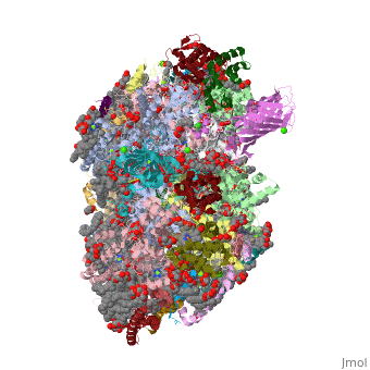
3wu2
From Proteopedia
Crystal structure analysis of Photosystem II complex
Structural highlights
Function[PSBL_THEVL] This protein is a component of the reaction center of photosystem II (PSII). PSII is a light-driven water plastoquinone oxidoreductase, using light energy to abstract electrons from H(2)O, generating a proton gradient subsequently used for ATP formation.[HAMAP-Rule:MF_01317] [YCF12_THEVL] A core subunit of photosystem II (PSII). PSII is a light-driven water plastoquinone oxidoreductase, using light energy to abstract electrons from H(2)O, generating a proton gradient subsequently used for ATP formation. [PSBB_THEVL] This protein binds multiple antenna chlorophylls and is part of the core of photosystem II (PSII). PSII is a light-driven water plastoquinone oxidoreductase, using light energy to abstract electrons from H(2)O, generating a proton gradient subsequently used for ATP formation. [PSBA_THEVL] D1 (PsbA) and D2 (PsbD) bind P680, the primary electron donor of photosystem II (PSII) as well as electron acceptors. PSII is a light-driven water plastoquinone oxidoreductase, using light energy to abstract electrons from H(2)O, generating a proton gradient subsequently used for ATP formation.[HAMAP-Rule:MF_01379] [PSBX_THEVL] Involved in the binding and/or turnover of quinones at the Q(B) site of photosystem II (PSII). PSII is a light-driven water plastoquinone oxidoreductase, using light energy to abstract electrons from H(2)O, generating a proton gradient subsequently used for ATP formation.[HAMAP-Rule:MF_01386] [CY550_THEVL] Low-potential cytochrome c that plays a role in the oxygen-evolving complex of photosystem II (PSII). Binds to PSII in the absence of other extrinsic proteins; required for binding of the PsbU protein to photosystem II. In PSII particles without oxygen-evolving activity, maximal activity is restored only by binding of cytochrome c550, PsbU and the 33 kDa PsbO protein. PSII is a light-driven water plastoquinone oxidoreductase, using light energy to abstract electrons from H(2)O, generating a proton gradient subsequently used for ATP formation.[1] [2] [PSBO_THEVL] Part of the oxygen-evolving complex associated with photosystem II (PSII). PSII is a light-driven water plastoquinone oxidoreductase, using light energy to abstract electrons from H(2)O, generating a proton gradient subsequently used for ATP formation. [PSBF_THEVL] This b-type cytochrome is tightly associated with the reaction center of photosystem II (PSII). PSII is a light-driven water plastoquinone oxidoreductase, using light energy to abstract electrons from H(2)O, generating a proton gradient subsequently used for ATP formation.[HAMAP-Rule:MF_00643] [PSBC_THEVL] This protein binds multiple antenna chlorophylls and is part of the core of photosystem II (PSII). PSII is a light-driven water plastoquinone oxidoreductase, using light energy to abstract electrons from H(2)O, generating a proton gradient subsequently used for ATP formation. [PSBJ_THEVL] This protein is a component of the reaction center of photosystem II (PSII). PSII is a light-driven water plastoquinone oxidoreductase, using light energy to abstract electrons from H(2)O, generating a proton gradient subsequently used for ATP formation.[HAMAP-Rule:MF_01305] [PSBM_THEVL] This protein is a component of the reaction center of photosystem II (PSII). PSII is a light-driven water plastoquinone oxidoreductase, using light energy to abstract electrons from H(2)O, generating a proton gradient subsequently used for ATP formation.[HAMAP-Rule:MF_00438] [PSBT_THEVL] Seems to play a role in the dimerization of photosystem II (PSII). PSII is a light-driven water plastoquinone oxidoreductase, using light energy to abstract electrons from H(2)O, generating a proton gradient subsequently used for ATP formation.[HAMAP-Rule:MF_00808] [PSBU_THEVL] Stabilizes the structure of photosystem II (PSII) oxygen-evolving complex (OEC), the ion environment of oxygen evolution and protects the OEC against heat-induced inactivation. Requires cytochrome c-550 (PsbV) or OEC3 (PsbO) to bind to photosystem II (PSII). PSII is a light-driven water plastoquinone oxidoreductase, using light energy to abstract electrons from H(2)O, generating a proton gradient subsequently used for ATP formation.[3] [PSBI_THEVL] A component of the reaction center of photosystem II (PSII). PSII is a light-driven water plastoquinone oxidoreductase, using light energy to abstract electrons from H(2)O, generating a proton gradient subsequently used for ATP formation.[HAMAP-Rule:MF_01316] [PSBD_THEVL] D1 (PsbA) and D2 (PsbD) bind P680, the primary electron donor of photosystem II (PSII) as well as electron acceptors. PSII is a light-driven water plastoquinone oxidoreductase, using light energy to abstract electrons from H(2)O, generating a proton gradient subsequently used for ATP formation. D2 is needed for assembly of a stable PSII complex.[HAMAP-Rule:MF_01383] [PSBZ_THEVL] Controls the interaction of photosystem II (PSII) cores with the light-harvesting antenna. PSII is a light-driven water plastoquinone oxidoreductase, using light energy to abstract electrons from H(2)O, generating a proton gradient subsequently used for ATP formation.[HAMAP-Rule:MF_00644] [PSBK_THEVL] This protein is a component of the reaction center of photosystem II (PSII). PSII is a light-driven water plastoquinone oxidoreductase, using light energy to abstract electrons from H(2)O, generating a proton gradient subsequently used for ATP formation. [PSBE_THEVL] This b-type cytochrome is tightly associated with the reaction center of photosystem II (PSII). PSII is a light-driven water plastoquinone oxidoreductase, using light energy to abstract electrons from H(2)O, generating a proton gradient subsequently used for ATP formation.[HAMAP-Rule:MF_00642] Publication Abstract from PubMedPhotosystem II is the site of photosynthetic water oxidation and contains 20 subunits with a total molecular mass of 350 kDa. The structure of photosystem II has been reported at resolutions from 3.8 to 2.9 A. These resolutions have provided much information on the arrangement of protein subunits and cofactors but are insufficient to reveal the detailed structure of the catalytic centre of water splitting. Here we report the crystal structure of photosystem II at a resolution of 1.9 A. From our electron density map, we located all of the metal atoms of the Mn(4)CaO(5) cluster, together with all of their ligands. We found that five oxygen atoms served as oxo bridges linking the five metal atoms, and that four water molecules were bound to the Mn(4)CaO(5) cluster; some of them may therefore serve as substrates for dioxygen formation. We identified more than 1,300 water molecules in each photosystem II monomer. Some of them formed extensive hydrogen-bonding networks that may serve as channels for protons, water or oxygen molecules. The determination of the high-resolution structure of photosystem II will allow us to analyse and understand its functions in great detail. Crystal structure of oxygen-evolving photosystem II at a resolution of 1.9 A.,Umena Y, Kawakami K, Shen JR, Kamiya N Nature. 2011 May 5;473(7345):55-60. Epub 2011 Apr 17. PMID:21499260[4] From MEDLINE®/PubMed®, a database of the U.S. National Library of Medicine. See AlsoReferences
| ||||||||||||||||||||
Categories: Large Structures | Thermosynechococcus vulcanus | Kamiya, N | Kawakami, K | Shen, J R | Umena, Y | Calcium binding | Chloride binding | Electron transport | Formylation | Hydroxylation | Iron binding | Manganese binding | Membrane complex | Oxygen evolving | Photosynthesis | Photosystem | Psii | Thylakoid membrane | Transmembrane alpha-helix | Water splitting
