Art:Polio: A resolution to eradicate
From Proteopedia
Behind the Artwork and the Protein Structure
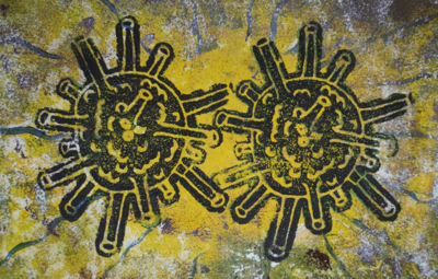
This artwork is an intaglio print of polio virus by Eric Lyle from Impington Village College. It is depicted in a space age style, and in the near future, we hope to have a world free from this disease. The near-eradication of poliomyelitis is one of the great success stories of modern medicine, but while polio might be almost eradicated, understanding the virus and how it infects us is still important. It’s likely that other viruses bind in a similar way to other receptors, starting their infectious cycle. Perhaps there are viruses similar to polio out there which could spontaneously mutate and become infectious to humans, so knowledge of how to combat them will be vital.
View in 3D or go to PDB structure 1dgi [1]
The poliovirus
Thanks to the efficacy of vaccination, poliomyelitis is now a very rare disease, one which the World Health Organisation have pledged to eradicate. The virus which causes it, poliovirus, has a football-shaped (icosahedral) shell formed from 60 copies of each of 3 different proteins. This arrangement is common to many viruses in the family of picornaviruses, including the one causing common cold.
In order to infect a human cell and replicate, poliovirus must first identify a cell and attach to it. It does this through interaction with a human protein called CD155, or poliovirus receptor (PVR). Despite its name, PVR normally sticks human cells together (cell adhesion), rather than sticking viruses to human cells. The polio vaccine triggers the body to make antibodies which coat the virus, preventing it from binding to PVR.
A low resolution view
Our first look at poliovirus’ interaction with PVR in the PDB archive came in 2000 from the Rossmann lab. Rossman used cryoelectron microscopy (cryoEM) to visualise the virus covered in PVR molecules. He could view the virus-PVR complex at 22Å resolution - not enough to see the position of atoms (which have a diameter of just under 2Å), but is still informative. It’s a bit like looking at a house from an aeroplane - you can see the overall shape and how big it is, but there’s no way you can make out the windows, or how the roof is tiled.
An important structure slowly revealed in beautiful clarity
Despite the low resolution of the PVR-bound structure, it could still be combined with other data to extract useful information. We already knew the detailed structure of poliovirus without PVR bound which was first solved by X-ray crystallography in 1989 (PDB entry 2plv), and structures of proteins similar to PVR were also known. With great care, a model of PVR bound to poliovirus could be built into the data (PDB entry 1dgi). It’s similar to attempting to model a house from an aerial photo, which is possible because you already know what windows look like, how bricks are arranged, and that front doors are usually on the ground floor. However, this house might, for example, have windows of a shape never seen before, so the model needs to be treated with caution and verified by how well it agrees with other experimental data.
Rossmann’s 22Å data was enough to confirm that poliovirus sticks to PVR using a ‘canyon’ on the virus surface. This is the same place where rhinovirus, which causes the common cold, latches on to its receptor. That 22Å data was obtained nearly 20 years ago. Amazing advances have been made in cryoEM since then which were recognised by the award of the Nobel Prize in 2017. These advances are reflected in the ever increasing resolution of structures of PVR/poliovirus solved by the technique.
By 2004, the complex was available at 15Å resolution (PDB entry 1nn8), and in 2015, the structure of poliovirus bound to PVR was solved to 3.7Å resolution PDB entry 3j8f. This is like moving from the aerial photo to a Google Street View of our house - maybe not quite good enough to see individual bricks in the walls, but if one or two were missing or sticking out, you could certainly tell! This structure of poliovirus showed exactly how PVR and the viral proteins interact, indicating that this interaction triggers changes in shape of the virus to enable the particle, and most importantly its genetic material, to enter the cell.
Explore the scientific publication
- ↑ He Y, Bowman VD, Mueller S, Bator CM, Bella J, Peng X, Baker TS, Wimmer E, Kuhn RJ, Rossmann MG. Interaction of the poliovirus receptor with poliovirus. Proc Natl Acad Sci U S A. 2000 Jan 4;97(1):79-84. PMID:10618374

