CHEM2052 Tutorial Example4
From Proteopedia
CHEM2052_Tutorial_Example4
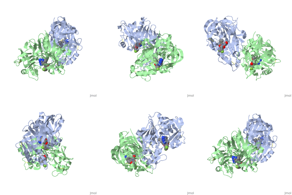
Chem2052: Example 4 - ReninThe spinning structure you initially view on this page is a protease called Renin (also known as angiotensinase). Renin is an aspartyl protease, which cleaves a particular peptide called angiotensinogen. Although this enzyme functions as a monomer the crystal structure obtained is a dimer and this is what you can see here. In the following representations one of the sub-units has been removed to represent the protein in its natural state. BackgroundIn later lecture we will look a little more closely at Renin. This enzyme is involved in a biological pathway leading to elevation of blood pressure, which can be beneficial in many ways. However if this process has become overactive, hypertension (high blood pressure) can result. Hypertension leads to cardiovascular disease which is the leading cause of death globally. The World Health Organisation states "An estimated 17.3 million people died from cardiovascular disease in 2008, representing 30% of all global deaths" see the following web page if you want to know more: Cardiovascular disease facts WHO. Since the 1970s scientists have been trying to modulate the action of renin by blocking the active site of the enzyme and preventing its function, hence lowering blood pressure. Aliskerin is the only renin inhibitor in clinical use today Renin information Site. However there is still interest in developing new, improved inhibitors. This question looks at a renin inhibitor identified through research at Pfizer.[1] Active SiteThis representation illustrates the of Renin. Consider the mechanism of the enzymatic cleavage of the natural peptide substrate by the catalytic residues you will find it here. Identify the structure of the transition state. In later lectures we will see how knowledge of this transition state can be used to design very effective enzyme inhibitors which mimic this structure, as you will find out, these are (not surprisingly) called "transition state" inhibitors. But for now though we will focus on the Pfizer inhibitor. Inhibition of ReninThis scene shows the . Which part of the inhibitor binds to the catalytic residues of the active site? Active Site InteractionsThis inhibitor does not just bind to the catalytic residues there are many other amino acids at the active site of the enzyme. First predict possible interaction types you would expect with this structure. Then look at the 2-dimensional "map" of the active site provided on your tutorial sheet and compare this to the 3-dimensional representations here. This view shows the amino acid residues on the of your "map". You should notice that these amino acids are fairly hydrophobic and interact with the hydrophobic parts of the inhibitor. If you prefer to view the structure where the inhibitor atoms are colour coded you will Similarly this view shows the amino acid residues on the of your "map". Again you can view this with the atoms of the inhibitor colour coded . References
| ||||||||||||

