Jmol/Cavities pockets and tunnels
From Proteopedia
| |||||||||||
Contents |
Preparing Isosurface Scenes for Proteopedia
Speed of Rendering
If you use the isosurface commands below, do so in the Jmol Java application, not in JSmol in Proteopedia. Depending on the size of the molecule, cavity isosurface commands take about a minute to complete in the Java application, which is many times faster than JSmol. You would have to wait many minutes for completion in JSmol.
Working Directory
Put a copy of Jmol.jar, and perhaps a downloaded copy of the PDB file(s) of interest in a new working directory (folder). By default, JVXL and PNGJ files you write will be written into the directory from which you start Jmol.jar. Any Jmol command script files should also be placed in this working directory. This keeps everything simple.
Use PNGJ Files
The best way to include cavity isosurfaces in a Proteopedia scene (green link) is to upload a properly prepared PNGJ file that includes the cached isosurfaces. Proteopedia's Molecular Scene Authoring Tools (SAT) load molecule tab accepts an uploaded PNGJ file.
Before saving the scene, you can add a caption that will display under the molecular display. The caption can include
- Links to pages within Proteopedia using double brackets, such as 6zgi (wikitext [[6zgi]]).
- Links to pages outside Proteopedia using single brackets, such as jmol.org (wikitext [http://jmol.org jmol.org]).
- Color keys using colored text. Coloring of text in captions must be done using span classes: see instructions at Coloring text. (In this example: <span class="text-red"><b>colored</b></span> <span class="bg-yellow">text</span>.)
Multiple Isosurfaces
There are two reasons why you may need multiple isosurfaces.
- You want to show both closed interior cavities and open-mouthed surface pockets.
- You want to show multiple pieces of a single isosurface.
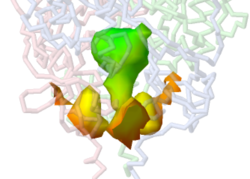
At present, Jmol can show only one piece of an isosurface. For example, above are shown three pocket entrances to the interior chamber of the spike protein cavity. Each pocket is displayed with an isosurface set <integer> command. But only one can be shown at a time. To display all 3, the JVXL file (see below) must be loaded 3 times, giving each a different ID, and then one pocket displayed from each. To also display the interior closed chamber, a 4th isosurface must be loaded. Below are scripts illustrating this.
Preparing PNGJ Files With Cached Isosurfaces
It is important to cache the isosurface(s) in your scene before saving the PNGJ file so the scene can load rapidly. If the PNGJ file is saved without caching, JSmol will re-calculate the isosurface(s) before the scene is displayed. This could easily take 10 minutes!
Here are the steps:
I. In the Jmol Java application:
- 1. Calculate the isosurface(s). Commands are explained above in earlier sections of this article. If you will need more than one isosurface, for example one for pockets and one for interior cavities, assign each a distinct ID when you create them. Prefixing the ID with tilde (~) ensures the ID name does not conflict with an existing term in Jmol. In a Jmol command line, anything after # is a comment, ignored by Jmol. For example:
- isosurface ~poc minset 100 pocket cavity
- isosurface ~bur minset 100 interior cavity # "buried"
- 2. Color the isosurface(s). Coloring by depth from the surface was described above. For a plain color, first you must know the ID of the isosurface if there are more than one.
- isosurface ~poc # Select the isosurface to color. Unnecessary if there is only one.
- color isosurface pink # color name
- isosurface ~bur # Select a different isosurface to color.
- color isosurface [x4060FF] # RGB color
- 3. Write each isosurface as a jvxl (Jmol Voxel) file. Coloring is saved in the JVXL file.
- isosurface ~poc # Select the isosurface to write. Unnecessary if there is only one.
- write poc.jvxl
- isosurface ~bur # Select a different isosurface to color.
- write bur.jvxl
- 4. Quit Jmol and start a new session. Load the molecule and then the JVXL file(s). If you are loading more than one JVXL file (or the same JVXL file more than once so you can display multiple pieces of it), assign each load a distinct ID. (ID's are not saved in the jvxl files.)
- load =6zgi
- isosurface ~poc1 poc.jvxl
- isosurface ~poc2 poc.jvxl
- isosurface ~poc3 poc.jvxl
- isosurface ~bur bur.jvxl
- 5. Cache the isosurface(s). This is critical. If this step is omitted, the PNGJ file will try to read the isosurfaces from the JVXL files, which will fail in Proteopedia.
- isosurface cache # A single command caches ALL isosurfaces.
- 6. Write the PNGJ file. It is best if you have rendered and colored the macromolecule exactly as you want the scene to appear in Proteopedia before writing the PNGJ file.
- write filename.pngj
II. In Proteopedia:
- 7. Upload your PNGJ file. You do NOT need to compress the PNGJ file because it is already in a compressed format. See Help:Uploading molecules. Keep a copy of the name of the page for your uploaded PNGJ file. For example, Image:6zgi-m1000-cav.pngj.
- 8. Now you are ready to use the SAT in your Proteopedia page. See Proteopedia:How to Make a Page. Use the load molecule tab in the SAT and paste in the name of the page containing your uploaded PNGJ file, such as Image:6zgi-m1000-cav.pngj. Use the save scene tab. Add a caption, and save the scene, creating a green link.
Working Example Scripts
The following script generates a pocket isosurface (open mouths) and an interior isosurface (closed pieces) for the spike protein, and saves each isosurface as a jvxl file. You can run this script: copy it and save it as a plain text file with a filename ending .spt. Drop the script file into the molecular display window of Jmol. Execution may take several minutes.
Write JVXL Files
# In the Jmol Java application,
# This script took 2 min 25 sec on a mid-2014 MacBook Pro.
# It would likely take ~20 min in JSmol.
zap # Deletes any molecule and all isosurfaces present in Jmol.
print # Clears the Jmol Script Console.

load =6zgi # Fetches PDB file from the RCSB Protein Data Bank.

# RENDER MOLECULE. Unnecessary but takes no time and looks nice while executing.
background white
rotate x -90
restrict protein # Selects protein and hides non-protein, e.g. glycosylation.
backbone only
backbone 0.7
color chain
set translucent false # Shows only the front-most surface of translucent objects.
color translucent 5 # Chain-colored backbones are now translucent.
refresh # Unnecessary, but shows the molecule.

# MAKE POCKETS
isosurface minset 50 pocket cavity 2.5 # Cavity probe radius 2.5 Å.
refresh
isosurface map property surfacedistance # Color pockets by depth.
refresh
write "poc.jvxl"

isosurface delete

# MAKE INTERIOR CAVITY NOT DISPLAYED IN THE POCKETS ISOSURFACE.
isosurface minset 100 interior cavity 2.5
refresh
isosurface map property surfacedistance
refresh
write "bur.jvxl" # Buried.
Write Cached PNGJ File
# Execution of this script took 4 seconds on a mid-2014 MacBook Pro.

zap # Deletes any molecule and all isosurfaces present in Jmol.
print # Clears the Jmol Script Console.

load =6zgi # Fetches PDB file from the RCSB Protein Data Bank.

# RENDER MOLECULE
background white
rotate x -90
restrict protein # Selects protein and hides non-protein, e.g. glycosylation.
backbone only
backbone 0.7
color chain
set translucent false # Shows only the front-most surface of translucent objects.
color translucent 5 # Chain-colored backbones are now translucent.
refresh # Unnecessary, but shows the molecule.

# DISPLAY OPEN POCKETS. Can display only one piece (set) per isosurface.
# To display three pieces (sets), create 3 (identical) isosurfaces.
# Set numbers determined by trial and error.
isosurface poc1 poc.jvxl
isosurface poc1 set 8

isosurface poc2 poc.jvxl
isosurface poc2 set 9

isosurface poc3 poc.jvxl
isosurface poc3 set 11

# CLOSED DEEPER CHAMBER.
isosurface bur bur.jvxl
isosurface bur set 1 # Unnecessary because set 1 is the only one passing minset 100.

delay 1.0 # shows the molecule with isosurfaces for 1 second.

# Arg1107 surrounds the deeper chamber.
# This centers that region, and zooms to 266%.
zoomto {arg1107} 266

isosurface cache # Without this, JSmol will fail trying to read the jvxl files absent in Proteopedia.
write "6zgi-pocbur2.5.pngj" # Ready to upload to Proteopedia.
Managing Cavity Isosurfaces
Jmol commands for generating cavity isosurfaces were explained above. Full details will be found in the Jmol/JSmol Interacive Scripting Documention under isosurfaces: molecular/solvent surfaces. Near the bottom of that very long section, important commands for after the cavity isosurfaces are calculated:
- isosurface area set (integer) # Reports the surface area of one piece (set) of an isosurface in Å2.
- isosurface * delete # Deletes all isosurfaces.
- isosurface set (integer) # Displays just one piece (set) of an isosurface.
- isosurface set 0 # Displays all pieces of an isosurface = the entire isosurface.
- isosurface volume set (integer) # Reports the volume of one closed[13] isosurface in Å3.
See Also
- Cavity programs
- PACUPP: Pockets And Cavities Using Pseudoatoms in Proteins
References and Notes
- ↑ Bondi, A., J. Phys. Chem. 68:441, 1964.
- ↑ Diameter of water molecule at B10NUMB3R5, a collaboration between Harvard Medical School and the Weizmann Institute of Science. See also Distance between water molecules in bulk water.
- ↑ Volume of water molecule bound to an amino acid at B10NUMB3R5, a collaboration between Harvard Medical School and the Weizmann Institute of Science.
- ↑ This command works only if a previous isosurface ... cavity command has been run in the same session. It will not work if the surface is loaded from a JVXL file alone, because then the surface has not been defined.
- ↑ The command for close examination is based on finding that Asn1107 is close to the deep chamber of the membrane-proximal cavity. The command is zoomto {1107} 266. This centers the deep part of the cavity and zooms to 266%.
- ↑ 6.0 6.1 Cavity of interest was manually disconnected from the largest cavity.
- ↑ Sum of volumes of cavity sets 5 and 11.
- ↑ Sum of volumes of 2 clusters of pseudoatoms.
- ↑ Sum of volumes of cavity sets 6, 9, 10, and 12.
- ↑ Sum of volumes of 2 clusters of pseudoatoms. One quarter of the cavity failed to be detected with a cavity probe radius of 2.5 Å.
- ↑ PACUPP in offset mode fails to find 3/4 of the cavity with a cavity probe radius of 2.5 Å.
- ↑ 12.0 12.1 Akama H, Kanemaki M, Yoshimura M, Tsukihara T, Kashiwagi T, Yoneyama H, Narita S, Nakagawa A, Nakae T. Crystal structure of the drug discharge outer membrane protein, OprM, of Pseudomonas aeruginosa: dual modes of membrane anchoring and occluded cavity end. J Biol Chem. 2004 Dec 17;279(51):52816-9. Epub 2004 Oct 26. PMID:15507433 doi:10.1074/jbc.C400445200
- ↑ The volume that Jmol reports for a pocket (open mouth/entrance) is meaningless. Jmol reports meaningful volumes only for closed isosurfaces.


