Structural and functional insights into phosphomannose isomerase
Mamata Bangera, Giri Gowda K., S.R. Sagurthi and M.R.N. Murthy [1]
Molecular Tour
Phosphomannose isomerase is a zinc binding enzyme that catalyses the reversible isomerization of mannose 6-phosphate and fructose 6-phosphate. These substrates could exist in two conformations. They are covalently closed (cyclic form) in one conformation while a covalent bond is disrupted in the other linear form. The reaction most likely proceeds by binding of the cyclic form of substrate, conversion of its closed to open form, transfer of protons between atoms of the open form of substrate by a suitable base followed by its cyclisation to form the cyclic form of the product.
Structure of phosphomannose isomerase from Salmonella typhimurium
The : an N terminal α-helical domain, a central catalytic domain and a C terminal domain. The carboxy, catalytic and helical domains are shown in red, green and blue, respectively. The polypeptide fold of the C terminal domain and catalytic domain are very similar to that of the cupin domain found in proteins belonging to different catalytic classes. The central catalytic domain contains the zinc binding site and has longer loops than the C terminal domain
Zinc binding site
. Mutation in any of these residues interferes with zinc binding, which in turn leads to a non-functional enzyme. Isothermal calorimetric experiments with mutants showed that loss of zinc binding is associated with lack of substrate binding as well. Hence zinc plays an important role in anchoring the substrate in the active site.
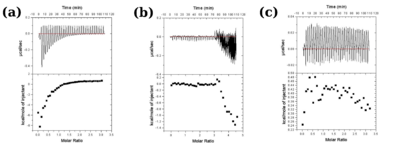
Figure 3: Isothermal titration calorimetric measurements for wild type protein (a), H99Q (b) and E134H (c) mutants. The characteristic profile observed for wild type protein indicative of binding of substrate is lost in the mutants.
Active site and catalytic base
Examination of . 
The colour coding represents conservation of sequence at residue positions, blue representing the least conserved and magenta representing the highly conserved positions. The active site pocket in the centre of the protein colocalises with the bound zinc ion (yellow sphere) and shows highest number of conserved residues.
. Zinc ion is shown as a grey sphere in the cupin domain (cartoon representation). Distance between Arg274 and the zinc ion is shown as a dashed line with the distance. In order to determine the most important residues for catalysis, site directed mutagenesis and activity studies were carried out on the mutants. Based on location in active site, conservation across species and complete loss of activity upon mutation, was proposed to be the catalytic base.
Concerted movement of residues
Lys132 and Arg274 were also identified to be important for the catalytic reaction by this and other studies. However, and the in the native enzyme. Comparison of structures of mutants with native enzyme revealed open and closed conformational states of the enzyme regulated by these residues. These states might be important for the binding of both the open and closed forms of the substrate and product in the catalytic cycle.
Two confomational states of StPMI. The loop (G116-H131) and helix (N101-115) occur in 2 orientations in wild type and mutant structures. Representative forms have been shown in red in and . .
PDB references: Phosphomannose isomerase from Salmonella typhimurium
References
- ↑ Bangera M, Gowda K G, Sagurthi SR, Murthy MRN. Structural and functional insights into phosphomannose isomerase: the role of zinc and catalytic residues. Acta Crystallogr D Struct Biol. 2019 May 1;75(Pt 5):475-487. doi:, 10.1107/S2059798319004169. Epub 2019 Apr 29. PMID:31063150 doi:http://dx.doi.org/10.1107/S2059798319004169


