Richards, Frederic M.
From Proteopedia
Frederic Middlebrook Richards (1925-2009[1][2][3]) was an eminent protein structure and function researcher[4][5], and a member of the US National Academy of Sciences. He earned his B.S. at MIT in 1948, and his Ph.D. at Harvard in 1952. He joined the faculty at Yale in 1955, where he became Chair of the Biophysics Department in 1963 and subsequently, in 1967, presided over the merger of Biophysics and Biochemistry into the Department of Molecular Biophysics and Biochemistry, where he became the Sterling Professor. At Yale, he led the creation of one of the major structural research centers in the world. Seven of the ten faculty he hired are now Members of the National Academy of Sciences[2]. His accomplishments include:
- Discovered, in the late 1950's, that inactive fragments of an enzyme could be recombined and regain activity, presaging Anfinson's work in the 1960's (Nobel Prize 1972), leading to the paradigm that amino acid sequence specifies the native conformation required for function. Richards' work was done with pancreatic ribonuclease A. He found that it could be proteolytically cleaved into a peptide and a protein fragment which, when separated were inactive, but could regain activity when properly recombined. This work began while Richards was a postdoc with Kaj Linderstrøm-Lang (Carlsberg Lab, Copenhagen) and continued after he joined the faculty at Yale.
- Solved the crystal structure of ribonuclease S in 1967 with faculty colleague Harold Wyckoff[6][7][8], including a structure with bound nucleoside monophosphate. This was the second enzyme structure solved, and the first protein crystallographic solution in the USA. See more about this structure below. A version of this structure was deposited in 1973 in the nascent Protein Data Bank (see below) as 1RNS[9] and is listed in the original PDB publication[10].
- Demonstrated that RNAse S was enzymatically active in crystalline form, strongly supporting the conclusion that the conformations of proteins in crystals are relevant to their native conformations in solution.
- Observed that the packing within a protein's core is as dense as organic molecules in molecular crystals such as sucrose[3]. This followed from his development of computational methods for analysis of protein packing.
- Developed, with B. K. Lee, computational methods for quantitating the solvent-accessible surfaces of proteins[1].

|
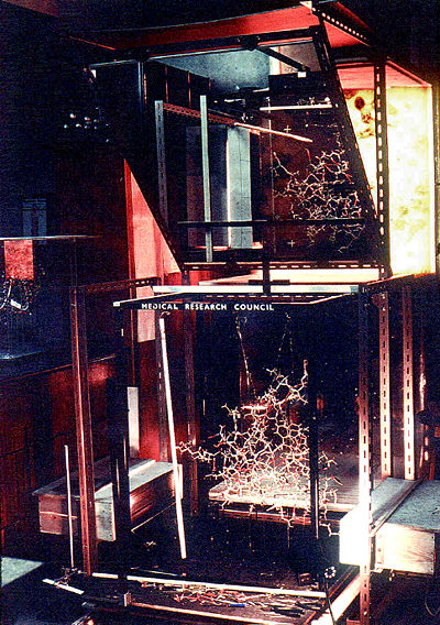
| The original optical comparator constructed by Fred Richards at Oxford in 1968. Behind the half-silvered mirror at the top is a transparent plastic sheet with a trace of a section of the electron density map. Gazing directly into the mirror, one can see the electron density superimposed with the physical model (at bottom). Light boxes at the bottom sides illuminate the section of the physical model represented by the electron density map. Multiple plastic sheets containing different sections of the electron density map are available in a rack at the top right (out of view). Photo given to User:Eric Martz in 2001 by Fred Richards with permission to share publicly. |
- Developed the optical comparator (see photo at right) in 1968 while on sabbatical at Oxford University[11]. Also known as the "Richards Box" or "Fred's Folly", this device enabled accurate manual fitting of a wireframe physical protein model to electron density maps by overlaying the two in a half-silvered mirror. With the convention, at the time, of building physical models at about one centimeter per Ångstrom, the device was taller than a person and filled the better part of a good sized room. It was widely adopted by crystallographers worldwide for the next decade, and the first computer-based replacements were initially called "electronic Richards boxes".
- Leading an early steering committee for the PDB, and a committee in the early 1980's that persuaded journal editors to require deposition of data in the PDB for structures reported in their journals, and the NIH to require data deposition as a condition for continued funding of research. These policies were established at a crucial time, just before a rapid increase in the rate at which macromolecular structural solutions were reported.[3] The Wyckoff and Richards strucure for RNAse S was deposited in the PDB in 1973 as 1RNS[9].
See Also
- 1RNS, the Wyckoff and Richards structure for RNAse S deposited in the PDB in 1973.
- Ribonuclease A, an article in the series Molecule of the Month.
- Ribonuclease A 1rta, with green links supplementing the article linked in the previous item.
References & Notes
- ↑ 1.0 1.1 Lim WA. Frederic M Richards 1925-2009. Nat Struct Mol Biol. 2009 Mar;16(3):230-2. PMID:19259121 doi:http://dx.doi.org/10.1038/nsmb0309-230
- ↑ 2.0 2.1 Steitz TA. Retrospective. Frederic M. Richards (1925-2009). Science. 2009 Feb 27;323(5918):1181. PMID:19251620 doi:http://dx.doi.org/323/5918/1181
- ↑ 3.0 3.1 3.2 Fox RO. Obituary: Frederic Richards (1925-2009). Nature. 2009 Feb 19;457(7232):976. PMID:19225517 doi:http://dx.doi.org/10.1038/457976a
- ↑ Richards FM. Whatever happened to the fun? An autobiographical investigation. Annu Rev Biophys Biomol Struct. 1997;26:1-25. PMID:9241411 doi:http://dx.doi.org/10.1146/annurev.biophys.26.1.1
- ↑ Staros, JV. Retrospective: Frederic M. Richards (1925-2009). ASBMB Today, April 2009, pp. 16-18.
- ↑ Wyckoff HW, Hardman KD, Allewell NM, Inagami T, Tsernoglou D, Johnson LN, Richards FM. The structure of ribonuclease-S at 6 A resolution. J Biol Chem. 1967 Aug 25;242(16):3749-53. PMID:6038503
- ↑ Wyckoff HW, Hardman KD, Allewell NM, Inagami T, Johnson LN, Richards FM. The structure of ribonuclease-S at 3.5 A resolution. J Biol Chem. 1967 Sep 10;242(17):3984-8. PMID:6037556
- ↑ Wyckoff HW, Tsernoglou D, Hanson AW, Knox JR, Lee B, Richards FM. The three-dimensional structure of ribonuclease-S. Interpretation of an electron density map at a nominal resolution of 2 A. J Biol Chem. 1970 Jan 25;245(2):305-28. PMID:5460889
- ↑ 9.0 9.1 Thanks to Jane Richardson for pointing out 1RNS
- ↑ Bernstein FC, Koetzle TF, Williams GJ, Meyer EF Jr, Brice MD, Rodgers JR, Kennard O, Shimanouchi T, Tasumi M. The Protein Data Bank: a computer-based archival file for macromolecular structures. J Mol Biol. 1977 May 25;112(3):535-42. PMID:875032
- ↑ Richards FM. The matching of physical models to three-dimensional electron-density maps: a simple optical device. J Mol Biol. 1968 Oct 14;37(1):225-30. PMID:5760491
Keywords
Biography, Obituary.
