SV40 Capsid Simplified
From Proteopedia
|
- (shows capsid only)
This model is based on the crystallographic solution of the VP1 capsid protein of Simian Virus 40, published as 1sva in 1996[1]. SV40 is a member of a group of cancer-causing viruses that has been extensively researched for decades.
The asymmetric unit of 1sva contains a pentamer of the VP1 protein plus one extra chain. In about 2010, the entire capsid was constructed from 1sva by the (now defunct) Probable Quaternary Structure server (of the European Bioinformatics Institute) using symmetry operations specified in 1sva.pdb (REMARK 350). The resulting capsid model contains 360 chains of VP1 protein arranged as 72 pentamers in an icosahedron (12 vertices forming 20 equilateral triangle faces). The PDB atomic coordinate file for the capsid, containing all non-hydrogen protein atoms, is 70 megabytes in size with about one million atoms, and 123,420 alpha carbon atoms.
In the highly simplified model at the right, each protein chain was reduced to a single atom, resulting in 360 atoms[2]. The distances between atoms were then greatly reduced to enable early versions of Jmol (and other common molecular visualization programs) to display the model more easily, with overlapping "atomic" spheres to simulate the capsid.
Yellow-colored are the twelve 5-chain pentamers representing the 12 vertices that define the 20 equilateral triangle faces of the icosahedron. Pseudoatoms were placed over each vertex to enable drawing lines representing the orientation of the icosahedron. Notice that each yellow 5-chain pentamer is surrounded by five pentamers, while each gray pentamer is surrounded by six pentamers.
Less-simplified capsid
FirstGlance in Jmol, since August, 2022, automatically constructs the capsid from the asymmetric unit of 1sv4, and automatically simplifies it to < 25,000 atoms to enable JSmol to handle it smoothly.
See less-simplified SV40 capsid in FirstGlance in Jmol.
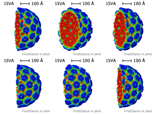
In the Views tab of FirstGlance, click Slab, and then check both of the checkboxes in the Slab options. You will get a result similar to the animation below. Can you pick out vertices with 5 and others with 6 nearest neighbors?
|
Half of the SV40 capsid, simplified by FirstGlance in Jmol, colored farthest from center and closest to center. Only every 5th alpha carbon is shown (24,720 alpha carbons). Atom diameters are exaggerated to make a solid-looking object. FirstGlance also offers the option of showing every 2nd alpha carbon (61,720 atoms) for a more detailed view. (Click the less simplified capsid link above, then under Biological Unit 1, click the link Show more detail.) |
Content Attribution
The original 2010 contents of this page were adapted from a site developed for Chime in the Atlas of Macromolecules with the permission of User:Eric Martz.
Notes
- ↑ Stehle T, Gamblin SJ, Yan Y, Harrison SC. The structure of simian virus 40 refined at 3.1 A resolution. Structure. 1996 Feb 15;4(2):165-82. PMID:8805523
- ↑ Since each original chain of 1sva had ~2,700 non-hydrogen atoms, this reduced the size of the data by ~2,700 fold
See Also
- The atomic coordinate file Image:Sv40 capsid 360 atoms.pdb. The capsid atoms are "chain V"; the icosahedron atoms are "chain I"; and the vertex atoms (yellow above) can be selected as "select 2". Pseudobonds were generated between the vertex atoms in Jmol with "connect 2 (:i) (:i)".

