Sandbox8999
From Proteopedia
|
Clathrin originates from the Latin word clāthrātus, meaning “to provide with a lattice”.[1] Clathrin is a protein involved in receptor-mediated endocytosis.[2] The protein was discovered by Barbara Pearse, a biological scientist, in 1975.[3] Clathrin is a protein resembling a triskelion shape and is composed of three heavy chains and three light chains which come together to form a polyhedral lattice similar to a cage.[2] The three heavy chains resemble three legs protruding from a center point. Some of the major functions of clathrin include lysosomal targeting, receptor-mediated endocytosis, and organelle biogenesis from the trans-Golgi network.[4] The polyhedral lattice shape of clathrin largely determines its functionality, in that there are many binding sites for proteins on the heavy chains of the lattice as well.[5]
Contents |
Structure
Individual Molecule
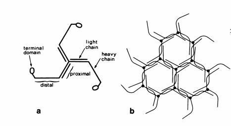
An individual clathrin molecule is composed of This structure is often referred to as a triskelion shape.[2] The heavy chains of clathrin are made of an amino-terminal, a beta-propellor domain which contain multiple repeats of the WD40 binding motif, alpha-helical zig-zags close to 30 amino acids in length, a lengthened alpha-helix where the threefold contacts, and a [2] The aforementioned zig-zags are what create the “leg” looking structure of the triskelion.[2] A segment of the heavy chain is designed with the purpose of attaching to the light chain.[2] The heavy chain and light chain bind through one long alpha helix towards the center of the protein.[2]

Lattice
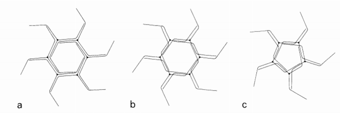
Individual clathrin molecules can assemble into cage-like structures. When individual clathrin molecules come together, a lattice is formed where each lattice point is associated with the center of a triskelion (from the Greek word meaning “a three legged structure”) (Figure 1).[2] According to Crowther, R.A., clathrin vesicles are constructed from twelve pentagonal units plus a number of hexagonal units, with the number of hexagons increasing for larger clathrin coats.[6] The term “cage” can be used to refer to empty shells composed only of clathrin while the term “coat” refers to clathrin in addition to associated proteins and vesicles.[6] Clathrin lattices are formed under optimal conditions of physiological pH (6.0-6.5) and in the presence of Calcium or Magnesium ions.[6] Various packing models have been investigated as to how the individual triskelion legs assemble into clathrin cages.[6] As mentioned above, the center of the triskelion legs occurs at each vertex of the cage, meaning pentagons, heptagons, and hexagons can be formed.[6] In the individual triskelion legs, the bend in the leg is 160 Angstroms from the vertex, giving rise to two models of packing: simple side-by-side packing or a cross-over type of packing (Figure 2).[6] Cross-over packing occurs when a hexamer of triskelions is formed with crossing over of the leg portions of the triskelions.[6] Rather than involving crossing over, simple packing refers to side by side packing of the leg portions of the triskelions (Figure 2).[6]

Function
Endocytosis
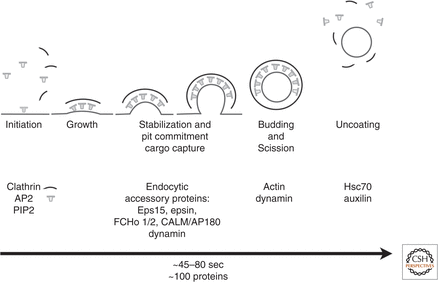
There are various endocytic mechanisms by which eukaryotic cells bring proteins and lipids into the cell, however clathrin-mediated endocytosis is known to be the most informative pathway to date.[7] Other portals for endocytic entry include phagocytosis and macropinocytosis.[7] Endocytic events are initiated through the activity of the clathrin coat and adaptor proteins that select the “cargo” that will be carried into the cell in vesicles.[8] At least twenty clathrin adaptors have been identified, and they all recognize and bind to membrane proteins or phospholipids that are specific for a certain organelle.[8] The formation of most cellular vesicles are facilitated by these coat-proteins like clathrin.[8] Cells endocytose vesicles from the plasma membrane to perform housekeeping tasks such as taking up nutrients, importing signaling receptors, mediating immune responses, cleaning up cell debris, or providing a pathway for pathogens or toxins.[8] Clathrin uses two main pathways for endocytosis: the canonical pathway and noncanonical pathway. The canonical clathrin pathway is referred to as the clathrin coated pits and the coated vesicles of endocytosis, whereas the noncanonical clathrin pathway is related to the clathrin-actin assembles of phagocytotic processes.[2]
The Canonical Pathway
Classical endocytosis involving clathrin-coated pits and clathrin-coated vesicles is referred to as the canonical clathrin pathway (Figure 3).[2] Canonical clathrin-dependent pathways involve the uptake of transferrin (Tf) by transferrin receptor (TfR) and the uptake of LDL by LDL receptors (LDLR).[2] Early studies showed that LDL epidermal growth factor (Tf) bound to their specific receptors could be endocytosed within the same vesicle.[2] The receptors for these molecules are composed of tails on their cytoplasmic side that contain sequences, which determine cargo sorting and also signal for clathrin to bind.[2] Clathrin then coats these vesicles and endocytosis occurs.[2] Other pathways, such as reuptake of neurotransmitters into the synaptic cleft, are seen to have similar mechanisms to canonical clathrin pathways.[2]

The Noncanonical Pathway
The noncanonical clathrin pathway refers to clathrin-actin assemblies, which form phagocytotic vesicles in response to invading bacterium.[2] Pathogen invasion into nonphagocytic cells result in the recruitment of clathrin.[2] These pathogens invade the cell through a process called the “zippering” mechanism, in which actin filaments are utilized to surround the pathogens with the host-cell membrane.[2] Actin recruitment requires the formation of clathrin patches and thus the clathrin-assembly forms.[2] Actin is not usually needed in classical endocytosis, but it is required when the size of the cargo in the forming vesicle is too large for the vesicle to close.[2] This association between actin and clathrin is unique to this pathway and distinguishes the noncanonical pathway from the canonical pathway.[2]
References
- ↑ Clathrin. (2015). Oxford University Press. Retrieved from http://www.oxforddictionaries.com/us/definition/american_english/clathrin.
- ↑ 2.00 2.01 2.02 2.03 2.04 2.05 2.06 2.07 2.08 2.09 2.10 2.11 2.12 2.13 2.14 2.15 2.16 2.17 2.18 2.19 2.20 2.21 Harrison, S. C., Kirchhausen, T., & Owen, D. (2014). Molecular structure, function, and dynamics of clathrin-mediated membrane traffic. Cold Spring Harbor Perspectives in Biology. doi: 10.1101/cshperspect.a016725
- ↑ 3.0 3.1 Pearse BM. Clathrin: a unique protein associated with intracellular transfer of membrane by coated vesicles. Proc Natl Acad Sci U S A. 1976 Apr;73(4):1255-9. PMID:1063406
- ↑ Wakeham, D. E., et al. (2003). Clathrin self-assembly involves coordinated weak interactions favorable for cellular recognition. The Embo Journal. doi: 10.1093/emboj/cdg511
- ↑ Ungewickell, E., & Brandon, D. (1981). Assembly units of clathrin coats. Nature, 289, 420-42. doi: 10.1038/289420a0
- ↑ 6.00 6.01 6.02 6.03 6.04 6.05 6.06 6.07 6.08 6.09 6.10 6.11 6.12 Crowther RA, Pearse BM. Assembly and packing of clathrin into coats. J Cell Biol. 1981 Dec;91(3 Pt 1):790-7. PMID:7328122
- ↑ 7.0 7.1 McPherson, P. S., Ritter, B., & Wendland, B. Clathrin-Mediated Endocytosis. (2009). Clathrin-Mediated Endocytosis. In Trafficking Inside Cells: Pathways, Mechanisms, and Regulation (Section II). doi: 10.1007/978-0-387-93877-6_9
- ↑ 8.0 8.1 8.2 8.3 Johnson, G. T., & Goodsell, D. (2007). Molecule of the Month: Clathrin. doi: 10.2210/rcsb_pdb/mom_2007_4
