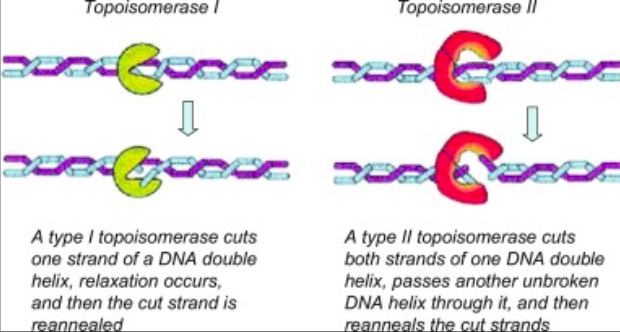
Eukaryotic DNA topoisomerase I (topo I) is a protein that reduces the strain from the supercoils that are caused during transcription and translation[1]. There are two types of topoisomerases. Type 1 topoisomerases are monomeric and break one strand of DNA[2]. Type 2 topoisomerases are dimeric, meaning that they made up of two units and break both strands of the DNA helix[2]. They are able to pass another part of the duplex through the cut, and close the cut using ATP[1].
 . [3].
Structure
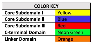
Human topo 1 is composed of 765 amino acids [2]. The enzyme consist of 4 regions which are the NH2-terminal, core, linker, and COOH-terminal domains[2]. The NH2-terminal is approximately 210 residues long, it is highly charged, disordered, and contains few hydrophobic amino acids[2]. The domain is made up of residues 713 to 765 and contains the important amino acid [2]. The location of the active site is at this amino acid[2]. Residues 636 to 712 form the and they contribute to the enzyme catalytic activity but are not required[2]. The core is divided into 3 subdomains: [215-232 & 320-433], [233-319], and [434-635][2]. The are very important for the catalytic activity[2].
Active Site & Mechanism Of Action
Topo 1 reduces stress in DNA by causing a transient single strand nick in the the DNA helix[1]. This nick enables the cut to rotate around its intact complement, thus eliminating proximal supercoils[1].
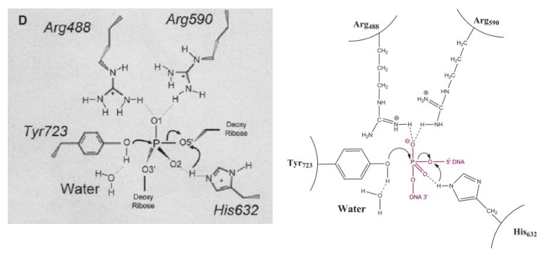
The active site of Topo 1 is catalytic and it is the location where the nicking or cutting occurs[2]. The nicking occurs from the trans-esterification of Tyr-723 at a DNA phophodiester bond forming a [1]. After the DNA is relaxed, the covalent intermediate is reversed when the released 5'-OH of the broken strand reattacks the phosphotyrosine intermediate in a second transesterification reaction[1].
 [4]
[4]
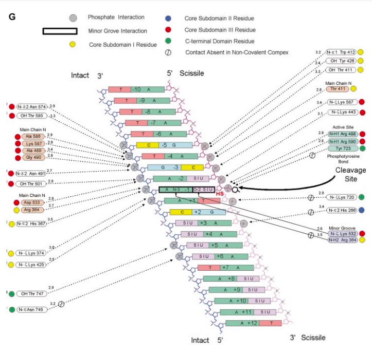 [4]
[4]
Relevance
Many anticancer drugs target topo 1 enzymes. This enzyme is the target of camptothecin (CPT) family of anticancer drugs[2]. These drugs work by increasing the duration of the nicked intermediate in the reaction [2]. CPT enhances DNA breakage at sites with a guanine base at the +1 position on the DNA strand that is being cut[2]. This is immediately downstream of the site that is being cleaved[2]. The stabilized intermediates prevent transcription and replication to continue in the cancer cells[2]. This eventually leads to DNA damage and cell death[2].
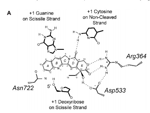 [2]
[2]
Mutations
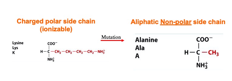
A mutation at amino acid 532 to Alanine almost abolishes enzyme activity[5]. The location of Lys 532 to the scissile phosphate and other active site amino acids could be the reason why a mutation of this amino acid abolishes the enzyme activity[5].
