Function of your protein
This enzyme, L-rhamnose- α-1,4-D-glucuronate lyase (FoRham1), is derived from the fungus Fusarium oxysporum and is a helpful tool for determining the structure and function of Gum Arabic (GA) to create potential agents to degrade GA more effectively. When the substrate GA is bound to FoRham1, the nonreducing ends of the glycosidic linkages are broken, releasing L-rhamnose (Rha) caps from GA [1]. Enzymes that can react with glycosidic linkages of certain carbohydrates can be useful in determining the structure, function, and mechanism of these carbohydrates, giving scientists the tools to manipulate their physical properties for further application to understand their breakdown. GA degrading specific enzymes are classified as glycoside hydrolases [1]. Determining this mechanism will give researchers a better understanding of how to degrade GA efficiently. Specifically, I focused on the mutant H105F, which has a PDB file of 7ESN.
Shown here is the enzyme using N->C coloring.
Biological relevance and broader implications
Gum Arabic (GA) is a representative protein of the family of arabinogalactan proteins (AGPs) and is produced in acacia trees in response to stress conditions, such as drought or wounds. GA has a variety of applications within the industrial world, including the food, cosmetic, and pharmaceutical industries, acting specifically as an emulsion stabilizer, emulsifier, and thickener in pharmaceutical settings. However, the the detailed structure of GA has not determined because of the complex branching that occurs in the polysaccharide. Enzymes that can react with and eliminate glycosidic linkages of carbohydrates are useful for determining the structure and function of these carbohydrates, giving researchers the opportunity to modify their physical properties. To date, there are no enzymes that have completely degraded GA [1].
Important amino acids
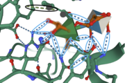
Amino Acids provide important interactions for binding in the active site. The amino acid residue is located near the C-5 atom of Rha, suggesting it has a catalytic role as a proton acceptor within the active site. Although there appears to be no catalytic triad in this protein, the side chain, which provides further stabilizing forces in the active site. Amino Acid Residues form a hydrogen bond with the O-1 atom of Rha, suggesting these residues also play a catalytic role for the elimination reaction. form hydrogen bonds with O-2 and O-3 atoms of Rha, providing further stabilization within the active site
[1].
The image to the right shows important interactions between the enzyme and amino acids in the active site. Hydrogen bonding and pi-stacking interactions are indicated by the blue and black circles, respectively.
Structural highlights
Secondary Structure: In this enzyme, there are around and three small . Compared to parallel beta sheets, anti-parallel beta sheets provide stronger hydrogen bonding between side chains of amino acids. The alpha helices provide structure for the formation of the active site, allowing the substrate (Rha) to bind in the active site.
Tertiary Structure: The structure of FoRham1 consists of a , providing a favorable conformation for the substrate binding into the active site, located on a central cleft towards the anterior side of the enzyme [1]. This beta-propeller is stabilized through hydrophobic interactions of the beta sheets, as well as hydrogen bonding that occurs between the N and C terminus of the beta sheets. B-propeller 1 is indicated by the red anti-parallel beta sheets, B-propeller 7 is indicated by the yellow anti-parallel beta sheets.
Quaternary Structure: This protein does not have quaternary structure.
Provided is the protein structure in space fill. This structure representation shows how much space certain molecules take up, indicating the depths of the active site and showing how deeply bound the ligand is to the enzyme. This representation also shows what size molecule fit into the active site, giving scientists an idea of other similar-sized ligands that may also fit into this binding pocket. Tan represents the enzyme, green represents the ligands, and blue represents the solvent.
Other important features
An is bound to the substrate complex, forming hydrogen bonds with amino acid residues Arg166 and Tyr202 [1]. The acetate ion provides more opportunity for the substrate to form interactions with amino acids near, or within, the active site, and may contribute to the elimination of the glycosidic linkages.
, I have provided a surface view of the substrate (Rha) bound into the enzyme. This view indicates how deep the cleft of the active site is as well as other areas within the structure. This view is important for indicating how other proteins or substrates interact with this enzyme. For example, you can use this view to get an idea if a substrate is too big to fit into the active site.

